. 2018 Sep 29;19(10):2981.
doi: 10.3390/ijms19102981.

Anthocyanins Extracted from Oryza sativa L. Prevent Fluorouracil-Induced Nuclear Factor-κB Activation in Oral Mucositis: In Vitro and In Vivo Studies

Affiliations

Anthocyanins Extracted from Oryza sativa L. Prevent Fluorouracil-Induced Nuclear Factor-κB Activation in Oral Mucositis: In Vitro and In Vivo Studies

Salunya Tancharoen et al. Int J Mol Sci. .

Abstract

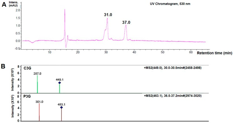
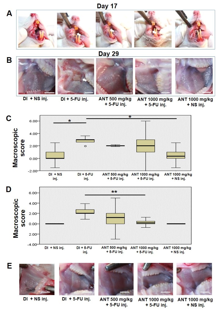
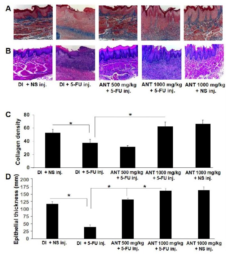
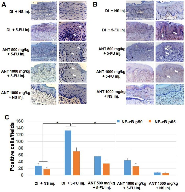
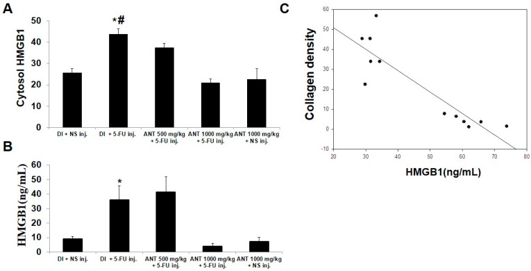
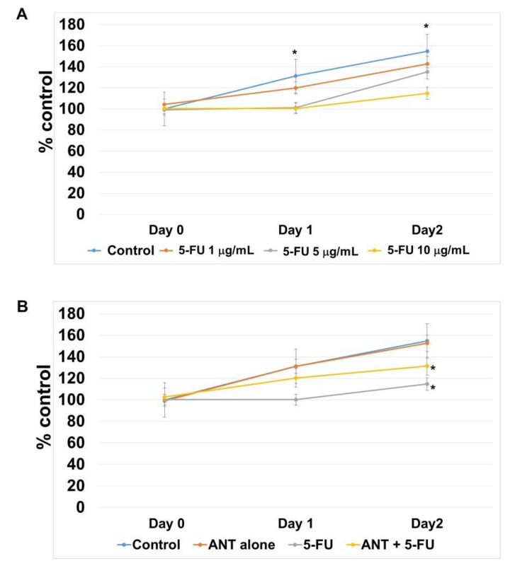
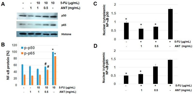
This study aims to investigate the immunomodulatory effect of anthocyanins (ANTs) from Oryza sativa L. extracts on 5-fluorouracil (5-FU)-induced oral mucositis, using a rat model and oral keratinocytes. ANTs were detected by high-performance liquid chromatography (HPLC)-electrospray ionization mass spectrometry. Animals were randomly given varying doses of ANT-rich extract treatment (500 mg/kg and 1000 mg/kg) in the absence or presence of 5-FU-induced mucositis. Buccal mucosae were photographed and scored for macroscopic analysis and incisional biopsies of cheek pouches were collected for microscopic examination of oral mucositis. 5-FU caused marked hemorrhage, extensive ulcerations and abscesses compared to non-treated animals with slight erythema. Histologically, a loss of collagen bundles and inflammatory cell infiltrates was observed. After 29 days of ANT treatment, lesions resolved, and abundant collagen fibers were evident in the lamina propria. Buccal mucosa of 5-FU-injected rats showed increased Nuclear factor-kappa B (NF-κB) p50 and p65 in oral keratinocytes. The administration of ANT reduced NF-κB-positive cells in 5-FU rats (p < 0.001) compared to the non-treatment group. In oral keratinocytes, ANT treatment significantly restored 5-FU-induced growth inhibition and impaired the nuclear accumulation of NF-κB p50 and p65. Our study demonstrated that ANT from Oryza sativa L. exhibited effective anti-inflammatory properties against 5-FU-induced oral mucositis by inhibiting NF-κB activation.

Keywords: 5-fluorouracil; NF-κB; anthocyanin; chemotherapy; oral mucositis.


Conflict of interest statement

The authors declare no conflict of interest.

Figures

Figure 1
Figure 1
HPLC-electrospray ionization (ESI) mass spectrometry (MS) analysis of anthrocyanins (ANTs) in Oryza sativa L. (A) Representative HPLC chromatograms of Cyanidin-3-glucoside (C3G) and Pelargonidin-3-glucoside (P3G) in the extracts at 530 nm. Peaks were detected with a retention time at 31.0 and 37.0 min, identified as C3G and P3G, respectively; (B) Fragmentation patterns in the mass spectra of C3G and P3G in the ANT. The assay was performed in triplicate.
Figure 2
Figure 2
Mean body weight of rats in different groups. Rats were gavage-fed with either deionized water (DI) or ANT-rich extract and injected with normal saline (NS) or 60 mg/kg of 5-fluorouracil (5-FU). Body weight was measured every two days. Data expressed as mean ± S.D. (n = 6 rats/group), * p < 0.05, vs. rats in the 5-FU treatment groups.
Figure 3
Figure 3
Pretreatment of rats with ANT-rich extract attenuates 5-FU-induced oral mucositis lesion. Rats were gavage-fed with either deionized water (DI) or ANT-rich extract and injected with normal saline (NS) or 5-FU. Macroscopic analysis of the oral mucositis lesion at the right buccal mucosa was observed on (A) day 17 and (B) day 29. Arrows indicate oral mucositis lesion. Scale bar, 10 mm; the box plots of the macroscopic scores of the mucositis lesion on (C) days 17 and (D) days 29 were measured by three examiners. Data are expressed as mean ± S.D., n = 6, Kruskall–Wallis test, * p < 0.001, ** p < 0.01. Box plots represent the median levels and the 25th and 75th percentiles of the observed data; whiskers represent the 5th and 95th percentiles in each group, and outliers (๐); (E) macroscopic analysis of the left buccal pouch without mechanical trauma on day 29. Scale bar, 10 mm.
Figure 4
Figure 4
Histological examination of buccal mucosa in 5-FU induced oral mucositis. (A) Mallory’s azan staining; (B) hematoxylin and eosin staining. Note that blue indicates collagen bundles stained by Mallory’s azan; original magnification ×40; (C) collagen density and (D) epithelial thickness were quantitated and data are expressed as mean ± S.D. * p < 0.001.
Figure 5
Figure 5
Localization of NF-κB p50 and p65 isoform in oral mucositis. By immunohistochemistry, slides were stained with (A) anti-NF-κB p50 Ab; (B) anti-NF-κB p65 Ab. Nuclei were counterstained with Mayer’s hematoxylin. Arrows indicate positive stained nuclei in oral epithelial cells. The arrowhead indicates inflammatory cells. Original magnification ×10 (left images) and ×40 (right images); (C) the number of NF-κB-positive cells per field. Data are expressed as the mean ± S.D. Error bars indicate standard deviation (n = 6), * p < 0.001.
Figure 6
Figure 6
The Evaluation of cytosolic and serum high mobility group box 1 (HMGB1) in 5-FU-induced oral mucositis. (A) Immunohistochemical analysis of cytosolic HMGB1 expression in the oral mucosa. The number of HMGB1-positive cells per field in the lesion have been count. * p < 0.05 vs. 500 mg/kg ANT-rich extract fed with 5-FU injection, # p < 0.001 vs. DI fed with normal saline (NS) injection; (B) serum HMGB1 levels were evaluated by ELISA. Data are expressed as the mean ± S.D. (n = 6). * p < 0.001 vs. 1000 mg/kg ANT-rich extract fed with 5-FU injection; (C) Correlation of the levels of collagen in the buccal mucosa and serum HMGB1.
Figure 7
Figure 7
The effect of ANT-rich extract on 5-FU-induced cell growth suppression. Viable cells were estimated by 3-[4,5-dimethylthiazol-2-yl]-2,5 diphenyl tetrazolium bromide (MTT) assay and expressed as percentage of control cells. (A) Cells were grown in medium supplemented with 5-FU (1, 5 and 10 μg/mL) for 2 days. 5-FU did not exert cytotoxic effects on oral keratinocytes. Data are expressed as the mean ± SD. * p < 0.05 vs. 5 and 10 μg/mL of 5-FU treatment; (B) cells were pretreated with 1 mg/mL ANT-rich extract and exposed to 5-FU (10 g/mL) for 2 days. * p < 0.05 vs. control.
Figure 8
Figure 8
Effects of ANT-rich extract on 5-FU-induced NF-κB phosphorylation. (A) Oral keratinocytes were stimulated with 5-FU in the presence or absence of ANT-rich extract for 1 h. Western blotting was performed for nuclear fraction of NF-κB p50 and p65, with histone as an equal loading control. The blots shown are representative of data from at least three experiments; (B) Quantifying nuclear NF-κB p 50 and p65 levels; data are expressed as the mean ± S.D. * p < 0.01 vs. control, # p < 0.05 vs. 1 mg/mL ANT-rich extract in the presence of 5-FU; the nuclear/cytoplasmic intensity ratio of (C) NF-κB p50 and (D) NF-κB p65. ANT-rich extract abundantly abrogated NF-κB nuclear translocation in response to 5-FU. Data are expressed as the mean ± S.D. * p < 0.05 compared to the 5-FU-treated cells.

Similar articles

Cited by

References

    1. Karthaus R.C., Ganser A. Prophylaxis and treatment of chemo-and radiotherapy-induced oral mucositis. Bone Marrow Transplant. 1999;24:1095–1108. doi: 10.1038/sj.bmt.1702024. - DOI - PubMed
    1. Sonis S.T. Mucositis as a biological process: A new hypothesis for the development of chemotherapy-induced stomatotoxicity. Oral Oncol. 1998;34:39–43. doi: 10.1016/S1368-8375(97)00053-5. - DOI - PubMed
    1. Logan R.M., Stringer A.M., Bowen J.M., Yeoh A.S., Gibson R.J., Sonis S.T. The role of pro-inflammatory cytokines in cancer treatment-induced alimentary tract mucositis: Pathobiology, animal models and cytotoxic drugs. Cancer Treat. Rev. 2007;33:448–460. doi: 10.1016/j.ctrv.2007.03.001. - DOI - PubMed
    1. Sonis S.T. Oral mucositis. Anti-Cancer Drugs. 2011;22:607–612. doi: 10.1097/CAD.0b013e3283462086. - DOI - PubMed
    1. Logan R.M., Stringer A.M., Bowen J.M., Gibson R.J., Sonis S.T., Keefe D.M. Is the pathobiology of chemotherapy-induced alimentary tract mucositis influenced by the type of mucotoxic drug administered? Cancer Chemother. Pharmcol. 2009;63:239–251. doi: 10.1007/s00280-008-0732-8. - DOI - PubMed