. 2018 Sep 30;19(10):2995.
doi: 10.3390/ijms19102995.

Prostaglandin E1-Mediated Collateral Recruitment Is Delayed in a Neonatal Rat Stroke Model

Affiliations

Prostaglandin E1-Mediated Collateral Recruitment Is Delayed in a Neonatal Rat Stroke Model

Philippe Bonnin et al. Int J Mol Sci. .

Abstract

While arterial reflow after a stroke represents an important challenge for better outcomes, it is also very important that sudden recanalization does not produce local oxidative and nitrogen species, deleterious for the brain and more particularly the immature brain. Our objective was to determine whether a supply in prostaglandin (Pg) E1 (Alprostadil), via its action on arterial pressure, might progressively improve cerebral reperfusion in a neonatal stroke model. Arterial blood flow was measured using ultrasonography. Rate-limiting and Pg terminal synthesizing enzymes were evaluated using reverse-transcriptase polymerase chain reaction. Our data suggests that a supply in PgE1 might delay and improve the ipsilateral reperfusion by decreasing thromboxane A synthase-1 gene, the density of reactive astrocytes and lesion volume.

Keywords: Astrocytes; Neonatal ischemia; hemodynamic responses; prostaglandins; thromboxane A synthase-1; ultrasound imaging.


Conflict of interest statement

The authors declare no conflict of interest.

Figures

Figure 1
Figure 1
Delayed thromboxane A synthase-1 (Tbxas) up-regulation after ischemia. (A) Gene expression for Cox-1 and Cox-2 rate limiting enzymes in sham (light grey bar) and PBS-treated ischemic animals at 1 (H1) and 2 (H2) hours after reperfusion in the contralateral (CL) and ipsilateral side (IL). (B) Gene expression for terminal prostaglandins synthesizing enzymes in sham and PBS-treated animals. * p <0.05 H2 vs. H1; # p < 0.05 H2 vs. Sham. (n = 6 at each time point in each group, and in sham (Sh) animals). mPGES1: microsomal PgE synthase-1; mPgES2: microsomal PgE synthase-2; PGIS: prostacyclin synthase.
Figure 2
Figure 2
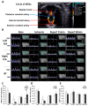
Blood-flow redistribution in the three great arteries of the circle of Willis after ischemia-reperfusion in P7 rats treated with PBS (n = 6) and/or prostaglandin E1 (PgE1) (n = 6). (A) Horizontal ultrasound cross-sectional imaging at the basis of the skull with color-Doppler mode showing the circle of Willis. The basilar trunk (BT) is revealed back, both intracranial internal carotid (ICA) and anterior cerebral arteries are revealed in red color at the front. The first branches of the ICAs, the posterior cerebral arteries are revealed in blue color (opposite flow-direction than in ICAs and BT). (B) After localization of the arteries, a pulsed-Doppler sample can be positioned on the different arteries for Doppler velocity waveforms recordings at the different steps of the protocol. (SP: systolic peak velocity; FD: end-diastolic velocity; MTMx: mean time maximal velocity; MTMn: mean time mean velocity; IP: index of pulsatility; IR: index of resistance; S/D systolic peak velocity/end-diastolic velocity. (C,D) is the mean blood flow velocities (mBFV) in the left and right ICAs decreased during ischemia; mBFVs recorded at 60 min of reperfusion remained lower than that measured in basal conditions in PBS-treated animals. In contrast, mBFVs returned to basal values in PgE1-treated rat pups (** p = 0.006, PgE1 vs. PBS). (E) mBFVs recorded in the BT showing no difference between PBS- and PgE1-treated animals. ** p < 0.01 PBS vs. PgE1.
Figure 3
Figure 3
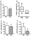
PgE1 induces better outcomes in neonatal ischemia at 48 h after ischemia-reperfusion. (A) Thromboxane A synthase-1 gene expression 2 h after ischemia-reperfusion in PBS- and PgE1-treated animals (n = 6 per group). (B) Infarct volumes in PBS- (n = 6) and PgE1-treated (n = 6) animals. The + indicates the mean. (C) Number of microglial cells and (D) GFAP density (astrocyte density) in the penumbra (n = 6 per group). * p < 0.05 vs. PBS; ** p < 0.01 vs. PBS.

References

    1. Wagenaar N., Martinez-Biarge M., van der Aa N.E., van Haastert I.C., Groenendaal F., Benders M.J.N.L., Cowan F.M., de Vries L.S. Neurodevelopment after perinatal arterial ischemic stroke. Pediatrics. 2018;142:e20174164. doi: 10.1542/peds.2017-4164. - DOI - PubMed
    1. Van Bel F., Dorrepaal C.A., Benders M.J., Zeeuwe P.E., van de Bor M., Berger H.M. Changes in cerebral hemodynamics and oxygenation in the first 24 h after birth asphyxia. Pediatrics. 1993;92:365–372. - PubMed
    1. Charriaut-Marlangue C., Bonnin P., Gharib A., Leger P.L., Villapol S., Pocard M., Gressens P., Renolleau S., Baud O. Inhaled nitric oxide reduces brain damage by collateral recruitment in a neonatal stroke model. Stroke. 2012;43:3078–3084. doi: 10.1161/STROKEAHA.112.664243. - DOI - PubMed
    1. Attwell D., Buchan A.M., Charpak S., Lauritzen M., Macvicar B.A., Newman E.A. Glial and neuronal control of brain blood flow. Nature. 2010;468:232–243. doi: 10.1038/nature09613. - DOI - PMC - PubMed
    1. Renolleau S., Aggoun-Zouaoui D., Ben-Ari Y., Charriaut-Marlangue C. A model of transient unilateral focal ischemia with reperfusion in the P7 neonatal rat: Morphological changes indicative of apoptosis. Stroke. 1998;29:1454–1461. doi: 10.1161/01.STR.29.7.1454. - DOI - PubMed

MeSH terms