. 2018 Nov 23;293(47):18180-18191.
doi: 10.1074/jbc.RA118.004652. Epub 2018 Oct 1.

Ligand binding and heterodimerization with retinoid X receptor α (RXRα) induce farnesoid X receptor (FXR) conformational changes affecting coactivator binding

Affiliations

Ligand binding and heterodimerization with retinoid X receptor α (RXRα) induce farnesoid X receptor (FXR) conformational changes affecting coactivator binding

Na Wang et al. J Biol Chem. .

Abstract

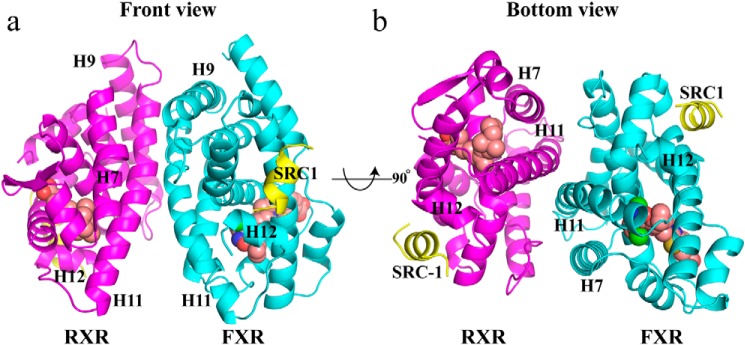
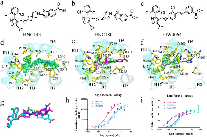
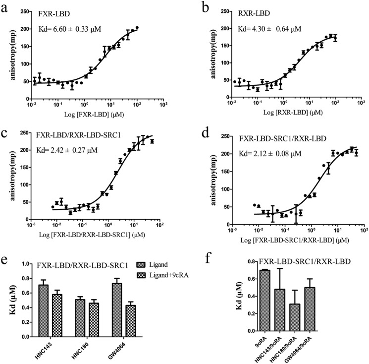
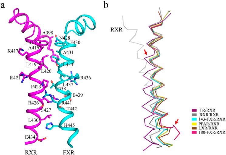
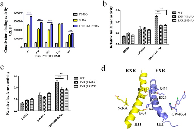
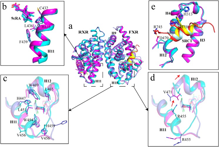
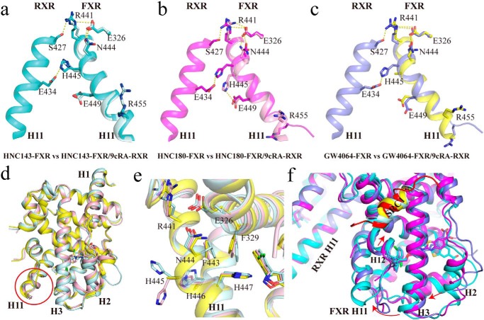
Nuclear receptor farnesoid X receptor (FXR) functions as the major bile acid sensor coordinating cholesterol metabolism, lipid homeostasis, and absorption of dietary fats and vitamins. Because of its central role in metabolism, FXR represents an important drug target to manage metabolic and other diseases, such as primary biliary cirrhosis and nonalcoholic steatohepatitis. FXR and nuclear receptor retinoid X receptor α (RXRα) form a heterodimer that controls the expression of numerous downstream genes. To date, the structural basis and functional consequences of the FXR/RXR heterodimer interaction have remained unclear. Herein, we present the crystal structures of the heterodimeric complex formed between the ligand-binding domains of human FXR and RXRα. We show that both FXR and RXR bind to the transcriptional coregulator steroid receptor coactivator 1 with higher affinity when they are part of the heterodimer complex than when they are in their respective monomeric states. Furthermore, structural comparisons of the FXR/RXRα heterodimers and the FXR monomers bound with different ligands indicated that both heterodimerization and ligand binding induce conformational changes in the C terminus of helix 11 in FXR that affect the stability of the coactivator binding surface and the coactivator binding in FXR. In summary, our findings shed light on the allosteric signal transduction in the FXR/RXR heterodimer, which may be utilized for future drug development targeting FXR.

Keywords: Farnesoid X receptor; Retinoid X receptor; X-ray crystallography; allosteric regulation; allostery; conformational change; metabolism; nuclear receptor; signal transduction; transcription factor.


Conflict of interest statement

The authors declare that they have no conflicts of interest with the contents of this article

Figures

Figure 1.
Figure 1.
FXR/RXRα heterodimer structure complexed with HNC143, 9cRA, and SRC1 peptides. a, ribbon diagram of the human FXR/RXRα heterodimer complex with two SRC1 peptides. The SRC1 peptides are in yellow. FXR is cyan, and RXRα is colored magenta. HNC143 in FXR and 9cRA in RXRα are shown in space-filling representation colored by atom type: oxygen as red, nitrogen as blue, sulfur as yellow, chlorine as green, and carbon as pink. b, heterodimer viewed from the bottom of H11, 90° rotation of a.
Figure 2.
Figure 2.
Chemical structure of the compounds and their interactions with FXR. a–c, the two-dimensional structures of HNC143, HNC180, and GW4064. d–f, ligand interactions with FXR–LBD in FXR/RXR heterodimer. The initial electron density maps calculated with σA-weighted FoFc coefficients, before the placement of ligands, are contoured at 3σ, 3σ, and 2σ, respectively. The proteins are represented as cartoons. The side chains of ligand-contacting residues (yellow for carbon, red for oxygen, blue for nitrogen, and green for sulfur) and the ligands (cyan for HNC143, magenta for HNC180, and slate for GW4064) are represented by sticks. Residues lining the binding pocket located within 4 Å of the ligands are shown. g, alignment of the three ligands in the heterodimer structures. h, dose-response curves of the FXR–LBD binding with SRC1 motif in the presence of HNC143, HNC180 and GW4064 as measured by AlphaScreen assays. RLU, relative light units. i, transcriptional activation assay of FXR–LBD with HNC143, HNC180, and GW4064. (EC50: HNC143, 0.306 μm; HNC180, 0.016 μm; GW4064, 0.159 μm.) 293T cells were transiently transfected with pCMV–GAL4–DBD–hFXR–LBD, PGL5, Renilla luciferase reporter plasmids. Relative activity was defined as pGL5–luciferase activity/Renilla luciferase activity. The data are shown as means ± S.D. All experiments were repeated at least three times.
Figure 3.
Figure 3.
Characterization of the interaction between coactivator and FXR–LBD, RXR–LBD, and FXR/RXR–LBD. a and b, binding curve of SRC1676–700 peptide to FXR–LBD, RXR–LBD monomer without ligands, measured by fluorescence anisotropy. c and d, binding curve of SRC1676–700 peptide to FXR/RXR–LBD·SRC1 and FXR–LBD·SRC1/RXR–LBD without ligands. The heterodimeric LBD complex consists of FXR–LBD and RXR–LBD fused with SRC1 or vice versa. This allows for the free biotinylated SRC1 peptide to bind only the unfused LBD. e and f, dissociation constants of the SRC1676–700 and FXR–LBD/RXR–LBD·SRC1 complexes (e) and FXR–LBD·SRC1/RXR–LBD (f), obtained from fluorescence anisotropy-based titrations. The experiments were carried out in the presence of FXR agonists (HNC143, HNC180, and GW4064), with or without RXR agonist (9cRA). The values are means ± S.D. of three independent experiments.
Figure 4.
Figure 4.
The FXR/RXRα heterodimer interface. a, intermolecular interactions mediated by the helices 11 of RXRα and FXR. Interacting residues on the heterodimer interface are labeled. b, alignment of different RXRα heterodimers and conformational variations of RXRα partner. Several NRs (colored as indicated) (TR/RXR: PDB code 4ZO1; PPAR/RXR: PDB code 1FM6; and LXR/RXR: PDB code 1UHL) were superimposed over the highly conserved N-terminal portion of helix 11 in RXR–LBD homodimer (PDB code 1MZN), corresponding to residues 413–427 in RXRα. TR and FXR H11 shifts are showed by red arrows.
Figure 5.
Figure 5.
The role of helix 11 in modulating the allosteric signal. a, FXR–LBD·SRC1/RXR–LBD (WT RXR–LBD and Glu434 mutants) proteins were used in coactivator recruitment assay, and FXR agonist GW4064 (1 μm) and RXR agonist 9cRA (1 μm) were added. RLU, relative light units. b and c, 293T cells were cotransfected with full-length WT FXR or FXR mutant expression plasmids, WT RXR or RXR mutant expression plasmids, FXR-responsive luciferase reporter (pGL3-IR1), and Renilla reporter plasmids. The cells were treated with DMSO, RXR agonist 9cRA (1 μm), and FXR agonist GW4064 (1 μm) for 24 h. Relative activity was defined as pGL3-luciferase activity/Renilla activity. The values are means ± S.D. of three independent experiments. **, p < 0.001; ***, p < 0.0001 (Student's t test). d, signal transduction pathway from the FXR/RXR heterodimer interface. H11 of FXR and RXR in GW4064-bound heterodimer are shown in cartoon; interacting residues on the heterodimer interface are shown in sticks.
Figure 6.
Figure 6.
The ligand-induced structural change in the C terminus of helix 11. a, alignment of the three FXR/RXR heterodimers. Cyan, heterodimer with HNC143; magenta, heterodimer with HNC180; slate, heterodimer with GW4064; pale cyan, SRC1 in HNC143 complex; red, SRC1 in HNC180 complex; yellow, SRC1 in GW4064 complex. b, shift in C terminus of H11 in RXR induced by heterodimerization with FXR and corresponding change in the position of RXR ligands 9cRA. c and d, the details of the difference on H11 and H12 with different ligands in FXR. e, coactivator binding interface (AF2 surface) of FXR in three heterodimers.
Figure 7.
Figure 7.
Ligand effect on the heterodimer interface and helix 11 conformation. a–c, overlay of FXR–LBD and FXR/RXR–LBD. Helix 10/11 is shown in pale cyan for HNC143–FXR–LBD and in cyan for HNC143–FXR/9cRA–RXR (a), in pink for HNC180–FXR–LBD and in magenta for HNC180–FXR/9cRA–RXR (b), and in yellow for GW4064–FXR–LBD and in slate for GW4064–FXR/9cRA–RXR(c). d, alignment of the three ligand-bound FXR–LBD monomer structures show the conserved C terminus of FXR H11. e, ligand-induced side-chain difference in H11 within three different ligand-bound monomeric FXR–LBD. f, the conformation change induced by the tail of FXR ligands. Alignment of three different FXR ligand-bound FXR/RXR heterodimers via superposition on RXR and colored as in a–c. Arrows show the shifts of H2, H3, H11, H12, and SRC1 in FXR.

References

    1. Lee F. Y., Lee H., Hubbert M. L., Edwards P. A., and Zhang Y. (2006) FXR, a multipurpose nuclear receptor. Trends Biochem. Sci. 31, 572–580 10.1016/j.tibs.2006.08.002 - DOI - PubMed
    1. Wang Y. D., Chen W. D., Moore D. D., and Huang W. (2008) FXR: a metabolic regulator and cell protector. Cell Res. 18, 1087–1095 10.1038/cr.2008.289 - DOI - PubMed
    1. Wang H., Chen J., Hollister K., Sowers L. C., and Forman B. M. (1999) Endogenous bile acids are ligands for the nuclear receptor FXR BAR. Mol. Cell 3, 543–553 10.1016/S1097-2765(00)80348-2,10.1016/S1097-2765(00)80484-0 - DOI - PubMed
    1. Fiorucci S., Cipriani S., Mencarelli A., Baldelli F., Bifulco G., and Zampella A. (2011) Farnesoid X receptor agonist for the treatment of liver and metabolic disorders: focus on 6-ethyl-CDCA. Mini. Rev. Med. Chem. 11, 753–762 10.2174/138955711796355258 - DOI - PubMed
    1. Darimont B. D., Wagner R. L., Apriletti J. W., Stallcup M. R., Kushner P. J., Baxter J. D., Fletterick R. J., and Yamamoto K. R. (1998) Structure and specificity of nuclear receptor-coactivator interactions. Genes Dev. 12, 3343–3356 10.1101/gad.12.21.3343 - DOI - PMC - PubMed

MeSH terms

Substances

LinkOut - more resources