. 2018 Dec 3;217(12):4284-4297.
doi: 10.1083/jcb.201805030. Epub 2018 Oct 1.

Bidirectional intraflagellar transport is restricted to two sets of microtubule doublets in the trypanosome flagellum

Affiliations

Bidirectional intraflagellar transport is restricted to two sets of microtubule doublets in the trypanosome flagellum

Eloïse Bertiaux et al. J Cell Biol. .

Abstract

Intraflagellar transport (IFT) is the rapid bidirectional movement of large protein complexes driven by kinesin and dynein motors along microtubule doublets of cilia and flagella. In this study, we used a combination of high-resolution electron and light microscopy to investigate how and where these IFT trains move within the flagellum of the protist Trypanosoma brucei Focused ion beam scanning electron microscopy (FIB-SEM) analysis of trypanosomes showed that trains are found almost exclusively along two sets of doublets (3-4 and 7-8) and distribute in two categories according to their length. High-resolution live imaging of cells expressing mNeonGreen::IFT81 or GFP::IFT52 revealed for the first time IFT trafficking on two parallel lines within the flagellum. Anterograde and retrograde IFT occurs on each of these lines. At the distal end, a large individual anterograde IFT train is converted in several smaller retrograde trains in the space of 3-4 s while remaining on the same side of the axoneme.


Figures

Figure 1.
Figure 1.
Positioning of IFT trains in the trypanosome flagellum and models for IFT trafficking. (A) Cross section of the trypanosome flagellum observed by conventional TEM. The arrowhead indicates an IFT particle positioned at the level of doublet 4. The cartoon shows the main structural components of the axoneme, with the numbering of microtubule doublets (Branche et al., 2006) superposed on the original image. Doublet numbering follows the conventional rules: a line perpendicular to the middle axis of the central pair microtubules is drawn and makes contact with the A tubule of only one doublet that is defined as number 1. The numbering follows the clockwise orientation defined by the dynein arms. Dynein arms are shown in orange, radial spoke in violet, central pair projections in yellow, and the PFR in blue. (B) The restricted presence of IFT on doublets 3–4 and 7–8 can be explained by two models: either some doublets are used specifically for anterograde transport and the other ones support retrograde IFT (Model 1) or bidirectional trafficking takes place on all doublets (Model 2). Only one doublet was highlighted for the sake of clarity.
Figure 2.
Figure 2.
IFT trains of similar length are distributed on doublets 3–4 and 7–8. (A) Successive images from Video 1 showing WT trypanosomes analyzed by FIB-SEM. Each image corresponds with a Z stack of three slices between positions 424 and 475. The progression is from anterior to posterior. Top: Low magnification of the cell body with major organelles indicated. F, flagellum; G, glycosome; M, mitochondrion. Bottom: A magnification of the flagellum area is shown with the axoneme (A) and the PFR. The arrows indicate IFT particles. (B) Portions of flagella reconstructed after FIB-SEM. Each axoneme is shown with a different color, and IFT trains are in red (for animation, see Video 2). (C) Another example of a flagellum from a WT trypanosome coming from a different stack than the one presented in A and B with the axoneme (sky blue) and several IFT trains (red). Doublet numbers and flagellum orientation (basal body and tip) are indicated. (D) Length of the IFT trains on doublets 3–4 (green; n = 52) and 7–8 (magenta; n = 56) determined from FIB-SEM analysis. Data are from 27 portions of flagella representing a total axoneme length of 166 µm. Two populations can be separated with short trains (green) and longer ones (magenta; see text for details).
Figure 3.
Figure 3.
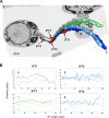
IFT trains are found closer to doublets 4 and 7 compared with doublets 3 and 8. (A) 3D view of a rare flagellum where individual doublets could be discriminated. Segmentation was performed to highlight the vicinity of the IFT trains (red) and microtubule doublets 3 (dark green), 4 (light green), 7 (light blue), and 8 (dark blue). (B) Plots representing the distances between the center of the skeleton for the indicated IFT trains and that of the microtubule doublets along their length using the same color code. In this flagellum, IFT trains are found closer to doublets 4 and 7.
Figure 4.
Figure 4.
IFT proteins are found on two distinct lines along the axoneme in fixed cells. (A and B) Control trypanosomes (strain expressing YFP::ODA8; Bonnefoy et al., 2018) were fixed in paraformaldehyde followed by methanol after fixation and processed for immunofluorescence using a marker antibody for the axoneme (middle; magenta on the merged image) and a monoclonal antibody against IFT172 (right; green on the merged image). The leftmost panel shows the phase-contrast image merged with DAPI staining (cyan). (A) Cell with one flagellum. (B) Cell assembling a new flagellum. In both cases, a single continuous thick line was observed for the axoneme marker, whereas discontinuous staining spreading on two close but distinct lines was visible for IFT172.
Figure 5.
Figure 5.
Bidirectional IFT trafficking takes place on two sides of the axoneme in live trypanosomes expressing mNG::IFT81. (A) Temporal projection of a stack of images corresponding with Video 5 showing the presence of two parallel lines for IFT in the flagellum in addition to the pool of IFT at the base. The left (green) and right (magenta) sides were defined after orientating the cell with the posterior end on top of the image and the flagellum on the left-hand side. (B) Still images from Video 5 of live trypanosomes expressing mNG::IFT81 imaged at high resolution. Green and magenta arrowheads indicate trains on the left and right sides, with light arrowheads indicating anterograde trains and darker arrowheads showing retrograde trains. The time point for each image is indicated. (C) Kymograph analysis of the same cell showing trafficking on the left side (green), on the right side (magenta), and the merged images for the region of interest indicated in A. Bars: 2.5 µm (horizontal bar); 2.5 s (vertical bar). (D) Dot plot of the frequency of anterograde IFT trains visible on the left (green) and the right (magenta) side, the sum of both (cyan), and from videos where only one track was visible (unresolved; gray). Values are coming from 18 different cells containing a total of 529 trains. *, P = 0.02. (E) Same representation but for the speed of anterograde trains. Only statistically significant differences are shown (one-way ANOVA test).
Figure 6.
Figure 6.
The IFT dynein travels on both sides of the axoneme in association with IFT proteins. (A) Temporal projection of the flagellum in a cell expressing GFP::DHC2.2. The region of interest (ROI) at the tip of the flagellum is indicated with left (green) and right (magenta) sides highlighted. (B) The kymographs are shown for the left and right side in the corresponding colors. Bars: 2.5 µm (horizontal bar); 5 s (vertical bar). (C) Kymograph analysis of a cell expressing mNG::IFT81 (left) and TdT::DHC2.1 (middle). On the merged panels, mNG::IFT81 is shown in green, and TdT::DHC2.1 is in magenta. Both proteins colocalized in moving but also in standing trains shown in the regions of interest. White arrowheads indicate arrested trains, while yellow arrowheads indicate an arrested train that started moving again. Even in these conditions, mNG::IFT81 and TdT::DHC2.1 remained associated. The white asterisk on the merged panel indicates a rare example of mNG::IFT81 material that was not associated with TdT::DHC2.1 and that remained immotile at the far distal end of the flagellum. However, the resolution was not sufficient to rule out the possibility that dynein could travel without cargo on other microtubules.
Figure 7.
Figure 7.
Anterograde trains are converted to retrograde trains while remaining on the same track. (A and B) IFT trafficking was analyzed in the GFP::IFT52-expressing cell line. The tip of the flagellum was bleached, and the arrival of new anterograde trains was monitored (see corresponding Video 10). For technical reasons, the bleaching event could not be recorded, and only the recovery phase can be presented. (A) Still images corresponding with the indicated times from Video 10. The thin green and magenta lines indicate the left and right sides of the axoneme, respectively. At 11 s, two anterograde trains were marked with a green (left side) or a magenta (right side) line, and the corresponding arrows indicate their direction. At 13 s, the left anterograde train converted to two retrograde ones that remained on the same left side of the flagellum. A second anterograde train arrived on the right side. At 14 s, the first left retrograde train left the field of view, and the second one progressed toward the base of the flagellum. The first anterograde train of the right side converted to a retrograde one that also remained on the right side. At 18 s, all retrograde trains on the left side were not in the field of view anymore as well as the first one on the right side. A new retrograde train was present on the right side and issued from the second anterograde train. Once again, conversion took place on the same side of the axoneme. (B) Kymograph analysis for the indicated left and right sides of the flagellum. The original kymograph is shown on the left of each panel, and the three annotated trains described above are marked in color.

Comment in

References

    1. Absalon S., Blisnick T., Kohl L., Toutirais G., Doré G., Julkowska D., Tavenet A., and Bastin P.. 2008. Intraflagellar transport and functional analysis of genes required for flagellum formation in trypanosomes. Mol. Biol. Cell. 19:929–944. 10.1091/mbc.e07-08-0749 - DOI - PMC - PubMed
    1. Adhiambo C., Blisnick T., Toutirais G., Delannoy E., and Bastin P.. 2009. A novel function for the atypical small G protein Rab-like 5 in the assembly of the trypanosome flagellum. J. Cell Sci. 122:834–841. 10.1242/jcs.040444 - DOI - PubMed
    1. Arganda-Carreras I., Fernández-González R., Muñoz-Barrutia A., and Ortiz-De-Solorzano C.. 2010. 3D reconstruction of histological sections: Application to mammary gland tissue. Microsc. Res. Tech. 73:1019–1029. 10.1002/jemt.20829 - DOI - PubMed
    1. Ball G., Demmerle J., Kaufmann R., Davis I., Dobbie I.M., and Schermelleh L.. 2015. SIMcheck: a Toolbox for Successful Super-resolution Structured Illumination Microscopy. Sci. Rep. 5:15915 10.1038/srep15915 - DOI - PMC - PubMed
    1. Bastin P., Sherwin T., and Gull K.. 1998. Paraflagellar rod is vital for trypanosome motility. Nature. 391:548 10.1038/35300 - DOI - PubMed

Publication types

MeSH terms