Review
. 2018 Oct 1;10(10):a032433.
doi: 10.1101/cshperspect.a032433.

The Story of RNA Folding, as Told in Epochs

Affiliations
Review

The Story of RNA Folding, as Told in Epochs

Daniel Herschlag et al. Cold Spring Harb Perspect Biol. .

Abstract

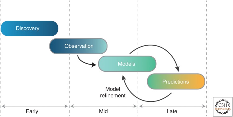
The past decades have witnessed tremendous developments in our understanding of RNA biology. At the core of these advances have been studies aimed at discerning RNA structure and at understanding the forces that influence the RNA folding process. It is easy to take the present state of understanding for granted, but there is much to be learned by considering the path to our current understanding, which has been tortuous, with the birth and death of models, the adaptation of experimental tools originally developed for characterization of protein structure and catalysis, and the development of novel tools for probing RNA. In this review we tour the stages of RNA folding studies, considering them as "epochs" that can be generalized across scientific disciplines. These epochs span from the discovery of catalytic RNA, through biophysical insights into the putative primordial RNA World, to characterization of structured RNAs, the building and testing of models, and, finally, to the development of models with the potential to yield generalizable predictive and quantitative models for RNA conformational, thermodynamic, and kinetic behavior. We hope that this accounting will aid others as they navigate the many fascinating questions about RNA and its roles in biology, in the past, present, and future.


Figures

Figure 1.
Figure 1.
Schematic of progress in a scientific field in terms of “epochs.”
Figure 2.
Figure 2.
Propensity of RNA to form base-pair interactions and stable secondary structure. (A) Short RNAs can form stable structures as a result of high stability of base-pair interactions as shown for the formation of a three-way junction. (B) The high stability of secondary structure results in formation of stable misfolded states with alternative secondary structures. Short peptides (green) can act as RNA chaperones and may have aided RNA function in the RNA World and provided a path from the RNA World to the RNA–protein world (Herschlag 1995).
Figure 3.
Figure 3.
Tetrahymena group I intron ribozyme and its P4-P6 domain. (A) Secondary structure of the Tetrahymena group I intron ribozyme. Long–range contacts are marked with dashed arrows. The P4-P6 domain is highlighted in red, with its metal core/metal core receptor (MC/MCR) and tetraloop/tetraloop receptor (TL/TLR) tertiary contacts colored in green and blue, respectively. Junction J5/5a (purple box) bends and allows coaxial stacks to be roughly parallel to each other. (B) X-ray structure of the P4-P6 domain with tertiary contacts colored as in A (PDB 1GID; Cate et al. 1996). (C) Electrostatic relaxation provided by cations followed by stabilization of a compact state by the formation of tertiary contacts. (D) Heterogeneous kinetic behavior of P4-P6 RNA caused by covalent damage during heating and ultraviolet (UV) irradiation. P4-P6 is shown in cartoon form with a pair of donor–acceptor fluorescent dyes (green and red stars) attached for single-molecule fluorescence resonance energy transfer (smFRET) folding studies. Scatter plots show values of the folding and unfolding rate constants (kF and kU, respectively) for individual P4-P6 RNA molecules prepared by a standard protocol (left) that involves heat/cool renaturation and UV irradiation and by a modified protocol (right) in which heating and UV radiation were eliminated during construct preparation and purification. (C, from Takamoto et al. 2004; reprinted, with permission, from Elsevier. D, from Greenfeld et al. 2011, adapted, with permission, from The American Society for Biochemistry and Molecular Biology.)
Figure 4.
Figure 4.
Kinetic and thermodynamic framework of the P4-P6 domain of the Tetrahymena group I intron (100 mm KCl, 5 mm MgCl2, pH 8, 23°C). The inset shows the overall folding rate constants observed in several studies of overall folding (for references, see Bisaria et al. 2016). (From Bisaria et al. 2016, modified, with permission.)
Figure 5.
Figure 5.
Hierarchical folding, sparsity of tertiary contacts, and structural modularity of RNA lead to a “reconstitution model” for RNA folding. (A) Schematic representation of a “reconstitution model” of RNA folding: (1) A complex RNA can be broken down into helix-junction-helix elements (HJH; cylinders and strings) and tertiary contact motifs (circles, squares, hexagons), and the energetic and conformational properties of these elements studied in isolation; (2) the behavior of the isolated elements can be reconstituted to understand their collective behavior in natural structured RNAs; and (3) novel RNAs can be constructed with structural motifs and can be engineered to have tunable energetic and conformational properties. (B) Free-energy diagram representing hierarchical folding of RNA. As an example, the folding of a simple RNA stabilized by a single tertiary contact is shown as a cartoon above the free-energy diagram. Helices are represented by cylinders, junctions as strings connecting the helices, and the single tertiary contact as a red square. (C) X-ray structures of the GAAA/11ntR tetraloop/tetraloop-receptor (TL/TLR) tertiary contact motif extracted from diverse structured RNAs and superimposed P4-P6 (PDB 1G1D; blue), RNase P (PDB 1NBS; green), and the Azoarcus group I intron (PDB 1ZZN; yellow and orange).
Figure 6.
Figure 6.
Schematic of a “reconstitution model” of RNA folding energetics. (A) Folding of a simple RNA involving the formation of a single tertiary contact (blue triangles) between two helices (cylinders) connected by a junction (strings). The free energy of folding (ΔGFold) is broken down into two terms: (1) the probability that the tertiary contact partners are found aligned for productive collision (ΔGAlign) and (2) the free energy associated with the formation of the tertiary contact (ΔGTert). (B) Each of the energy terms in A is represented in 3D free-energy landscapes. ΔGAlign can be further broken down into an energy term intrinsic to the conformational preferences of the junction and the helices containing the tertiary contact partners (ΔGHJH) and an energy term reflecting the electrostatic interactions between RNA and surrounding ions and between RNA helices (ΔG+/−). (C) Quantitative prediction of the “reconstitution model” of RNA. According to this model, the free energy of formation of a tertiary contact ΔGTert is separable from the free energies associated with the conformational search of the junction ΔGTert and the electrostatic interactions with ions ΔG+/−. Hence, this model predicts that the energetic effect of mutating a tertiary contact motif is constant across different ionic conditions. (From Bisaria et al. 2017, reprinted, with permission.)
Figure 7.
Figure 7.
Conformational rearrangements on tetraloop/tetraloop receptor (TL/TLR) formation. (A) Comparison of undocked (top) and docked (bottom) structures of the canonical GAAA/11ntR TL/TLR motif indicate that the 11ntR tetraloop-receptor undergoes a series of conformational changes on binding to the GAAA tetraloop (Cate et al. 1996; Butcher et al. 1997; Davis et al. 2005). The structure of the GAAA tetraloop does not change significantly on docking (Heus and Pardi 1991; Fiore and Nesbitt 2013). (B) Single-molecule fluorescence resonance energy transfer (smFRET) dissection of the folding kinetics and thermodynamics of a set of TL/TLR sequence variants across a range of ionic conditions led to a model for the order of conformational changes along the folding pathway of the TL/TLR motif (Bonilla et al. 2017). (From Bonilla et al. 2017, reprinted, with permission, from the American Chemical Society.)
Figure 8.
Figure 8.
RNA-MaP method applied to the study of RNA tertiary elements. (A) The RNA-MaP platform with a bound expressed RNA (“chip” piece) and a “flow” piece RNA added in trans (Denny et al. 2018). These RNAs combine to form a tertiary “TectoRNA.” (B) A model for the tertiary structure of the TectoRNA homodimer (PDB: 2ADT; Davis et al. 2005). A heterodimer in which one of the canonical GAAA/11ntR TL/TLRs is replaced by an orthogonal GGAA/R1 (Geary et al. 2008) was used as a “host” for different helix sequences and lengths, different junction elements, and different tertiary motif elements. (C) Examples of junction elements used in these studies. (A,C, Reproduced, with permission, from Denny et al. 2018.)

References

    1. Adamala K, Szostak JW. 2013. Nonenzymatic template-directed RNA synthesis inside model protocells. Science 342: 1098–1100. - PMC - PubMed
    1. Bailor MH, Sun X, Al-Hashimi HM. 2010. Topology links RNA secondary structure with global conformation, dynamics, and adaptation. Science 327: 202–206. - PubMed
    1. Bisaria N, Greenfeld M, Limouse C, Pavlichin DS, Mabuchi H, Herschlag D. 2016. Kinetic and thermodynamic framework for P4-P6 RNA reveals tertiary motif modularity and modulation of the folding preferred pathway. Proc Natl Acad Sci 113: E4956–E4965. - PMC - PubMed
    1. Bisaria N, Greenfeld M, Limouse C, Mabuchi H, Herschlag D. 2017. Quantitative tests of a reconstitution model for RNA folding thermodynamics and kinetics. Proc Natl Acad Sci 114: E7688–E7696. - PMC - PubMed
    1. Bonilla S, Limouse C, Bisaria N, Gebala M, Mabuchi H, Herschlag D. 2017. Single-molecule fluorescence reveals commonalities and distinctions among natural and in vitro-selected RNA tertiary motifs in a multistep folding pathway. J Am Chem Soc 139: 18576–18589. - PMC - PubMed

Publication types

LinkOut - more resources