. 2018 Oct 1;11(10):1880.
doi: 10.3390/ma11101880.

Encapsulation of Rat Bone Marrow Derived Mesenchymal Stem Cells in Alginate Dialdehyde/Gelatin Microbeads with and without Nanoscaled Bioactive Glass for In Vivo Bone Tissue Engineering

Affiliations

Encapsulation of Rat Bone Marrow Derived Mesenchymal Stem Cells in Alginate Dialdehyde/Gelatin Microbeads with and without Nanoscaled Bioactive Glass for In Vivo Bone Tissue Engineering

Ulrike Rottensteiner-Brandl et al. Materials (Basel). .

Abstract

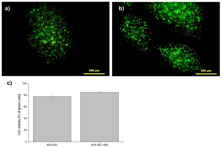
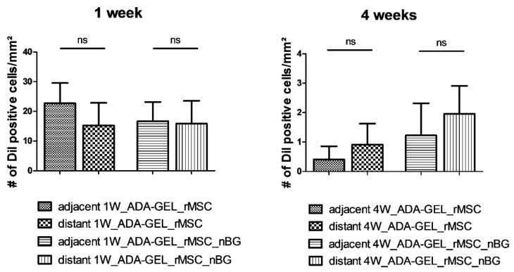
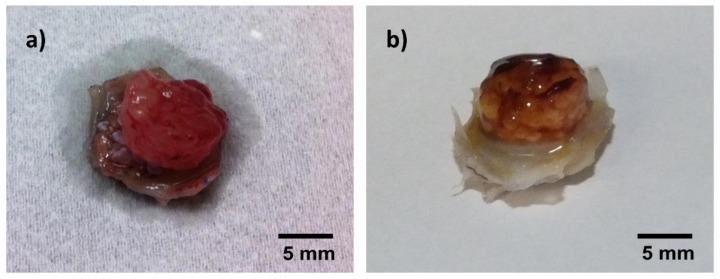
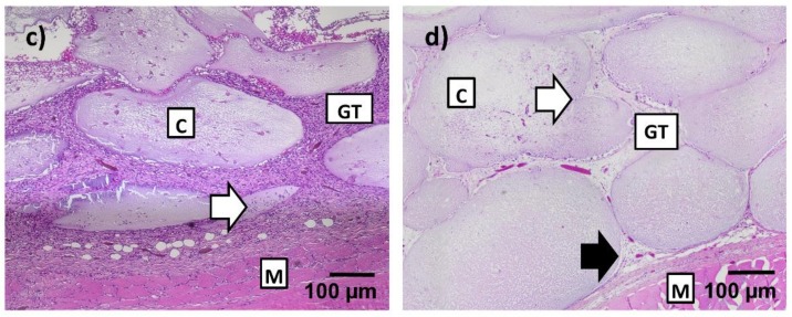
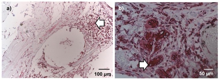
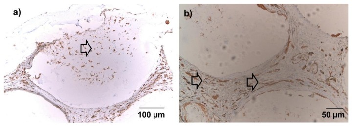
Alginate dialdehyde (ADA), gelatin, and nano-scaled bioactive glass (nBG) particles are being currently investigated for their potential use as three-dimensional scaffolding materials for bone tissue engineering. ADA and gelatin provide a three-dimensional scaffold with properties supporting cell adhesion and proliferation. Combined with nanocristalline BG, this composition closely mimics the mineral phase of bone. In the present study, rat bone marrow derived mesenchymal stem cells (MSCs), commonly used as an osteogenic cell source, were evaluated after encapsulation into ADA-gelatin hydrogel with and without nBG. High cell survival was found in vitro for up to 28 days with or without addition of nBG assessed by calcein staining, proving the cell-friendly encapsulation process. After subcutaneous implantation into rats, survival was assessed by DAPI/TUNEL fluorescence staining. Hematoxylin-eosin staining and immunohistochemical staining for the macrophage marker ED1 (CD68) and the endothelial cell marker lectin were used to evaluate immune reaction and vascularization. After in vivo implantation, high cell survival was found after 1 week, with a notable decrease after 4 weeks. Immune reaction was very mild, proving the biocompatibility of the material. Angiogenesis in implanted constructs was significantly improved by cell encapsulation, compared to cell-free beads, as the implanted MSCs were able to attract endothelial cells. Constructs with nBG showed higher numbers of vital MSCs and lectin positive endothelial cells, thus showing a higher degree of angiogenesis, although this difference was not significant. These results support the use of ADA/gelatin/nBG as a scaffold and of MSCs as a source of osteogenic cells for bone tissue engineering. Future studies should however improve long term cell survival and focus on differentiation potential of encapsulated cells in vivo.

Keywords: alginate dialdehyde; bioactive glass; gelatin; mesenchymal stem cells; nanoparticles; tissue engineering.


Conflict of interest statement

The authors declare no conflict of interest. The funders had no role in the design of the study; in the collection, analyses, or interpretation of data; in the writing of the manuscript, and in the decision to publish the results.

Figures

Figure 1
Figure 1
Fluorescence microscopy images of rat bone marrow derived mesenchymal stem cells (rMSCs) cells encapsulated in alginate dialdehyde-gelatin (ADA-GEL) (a) and ADA-GEL-nano-scaled bioactive glass (nBG), (b) after encapsulation (a,b: green—calcein-stained live cells in images, red—PI-stained dead cells; cells were incubated for 7 days before live-dead staining was performed). Quantitative analysis of cell viablity in ADA-GEL and ADA-GEL-nBG hydrogels, performed by image analysis (c).
Figure 2
Figure 2
(a) Representative image 1 week after implantation (1W_ADA-GEL_rMSC). DiI (DiI cell tracker, Life Technologies, Darmstadt, Germany) -labeled rMSCs (red) are easily detectable within beads, and their nuclei are clearly co-stained with DAPI (blue), proving their viability; (b) Mean total number of DiI positive cells per mm2 (mean ± SD). A marked decrease was noted after 4 weeks, compared to cell survival after 1 week. ns: not significant, ** p ≤ 0.01 (Mann Whitney U Test).
Figure 3
Figure 3
Mean number (+/− SD) of DiI positive cells per mm2 in different areas of the slide. Surprisingly, a higher number of DiI positive cells was present distant from the muscle after 4 weeks, compared to the areas adjacent to the muscle, although this difference was not statistically significant (ns) (Mann Whitney U Test).
Figure 4
Figure 4
Macroscopic and microscopic (hematoxylin-eosin (H&E)-stained) appearance of constructs after explantation, representative images (not all groups are depicted, as no macroscopic difference was noticed between constructs with and without nBG). (a) macroscopic image, Group 1W_ADA-GEL-rMSC-nBG; loosely adhering, moist, transparent hydrogel beads; (b) macroscopic image, Group 4W_ADA-GEL-rMSC; firm, well adhering, opaque hydrogel beads; (c) H&E staining, 10×, group 1W_ADA-GEL-rMSC; loose, highly cellular granulation tissue in the areas between beads, variable degree of infiltration with small cells (arrow); (d) H&E staining, 10×, group 4W_ADA-GEL_rMSC; thin septae of connective tissue infiltrating the capsules (white arrow); a thin gap is visible between capsules and connective tissue (black arrow); GT = granulation tissue, C = capsules, M = muscle.
Figure 4
Figure 4
Macroscopic and microscopic (hematoxylin-eosin (H&E)-stained) appearance of constructs after explantation, representative images (not all groups are depicted, as no macroscopic difference was noticed between constructs with and without nBG). (a) macroscopic image, Group 1W_ADA-GEL-rMSC-nBG; loosely adhering, moist, transparent hydrogel beads; (b) macroscopic image, Group 4W_ADA-GEL-rMSC; firm, well adhering, opaque hydrogel beads; (c) H&E staining, 10×, group 1W_ADA-GEL-rMSC; loose, highly cellular granulation tissue in the areas between beads, variable degree of infiltration with small cells (arrow); (d) H&E staining, 10×, group 4W_ADA-GEL_rMSC; thin septae of connective tissue infiltrating the capsules (white arrow); a thin gap is visible between capsules and connective tissue (black arrow); GT = granulation tissue, C = capsules, M = muscle.
Figure 5
Figure 5
ED1 (CD68) immunohistochemical staining after 4 weeks, 10× (a) and 20× magnification (b), representative images (not all groups are depicted). A low number of ED1 positive cells was present in the granulation tissue (a; arrow). Few foreign body giant cells were detected in single constructs (b; arrow). No differences were evident between groups with nBG and without nBG in qualitative microscopic evaluation.
Figure 6
Figure 6
Lectin immunohistochemical staining, 10× (a) and 20× magnification (b), representative images (not all groups are depicted). Brown cells = lectin-positive; blue cells = lectin-negative (counterstained with hemalaun). (a) After 1 week, lectin-positive cells were diffusely dispersed in the connective tissue between beads. A fraction of the infiltrating cells stained clearly positive for lectin (arrow). (b) After 4 weeks, the connective tissue between the microbeads was highly vascularized (arrows). No differences were evident between groups with nBG and without nBG in qualitative microscopic evaluation.
Figure 7
Figure 7
Quantitative evaluation/mean number (+/− SD) of lectin-positive cells per slide. Lectin counts from slides from a previous study with the same study design without encapsulated rMSCs [18] were used for comparison. Group 4W_ADA-GEL-rMSC showed a significantly higher number than group 4W_ADA-GEL, and group 4W_ADA-GEL-rMSC-nBG exhibited a higher number of lectin-positive cells than both groups without encapsulated cells. ns: not significant; * p ≤ 0.05; ** p ≤ 0.01 (ANOVA/Bonferroni’s Post Hoc).

References

    1. Drury J.L., Mooney D.J. Hydrogels for tissue engineering: Scaffold design variables and applications. Biomaterials. 2003;24:4337–4351. doi: 10.1016/S0142-9612(03)00340-5. - DOI - PubMed
    1. Andersen T., Strand B.L., Formo K., Alsberg E., Christensen B.E. Alginates as biomaterials in tissue engineering. Carbohydr. Chem. 2008;37:227–258.
    1. Duan B., Hockaday L.A., Kang K.H., Butcher J.T. 3D bioprinting of heterogeneous aortic valve conduits with alginate/gelatin hydrogels. J. Biomed. Mater. Res. Part A. 2013;101:1255–1264. doi: 10.1002/jbm.a.34420. - DOI - PMC - PubMed
    1. Rokstad A.M., Brekke O.L., Steinkjer B., Ryan L., Kollarikova G., Strand B.L., Skjak-Braek G., Lacik I., Espevik T., Mollnes T.E. Alginate microbeads are complement compatible, in contrast to polycation containing microcapsules, as revealed in a humand whole blood model. Acta Biomater. 2011;7:2566–2578. doi: 10.1016/j.actbio.2011.03.011. - DOI - PubMed
    1. Veriter S., Mergen J., Goebbels R.M., Aouassar N., Gregoire C., Jordan B., Leveque P., Gallez B., Gianello P., Dufrane D. In vivo selection of biocompatible alginates for islet encapsulation and subcutaneous transplantation. Tissue Eng. Part A. 2010;16:1503–1513. doi: 10.1089/ten.tea.2009.0286. - DOI - PubMed