. 2018 Oct 1;8(1):14567.
doi: 10.1038/s41598-018-32803-6.

Partial depletion of CD206-positive M2-like macrophages induces proliferation of beige progenitors and enhances browning after cold stimulation

Affiliations

Partial depletion of CD206-positive M2-like macrophages induces proliferation of beige progenitors and enhances browning after cold stimulation

Yoshiko Igarashi et al. Sci Rep. .

Abstract

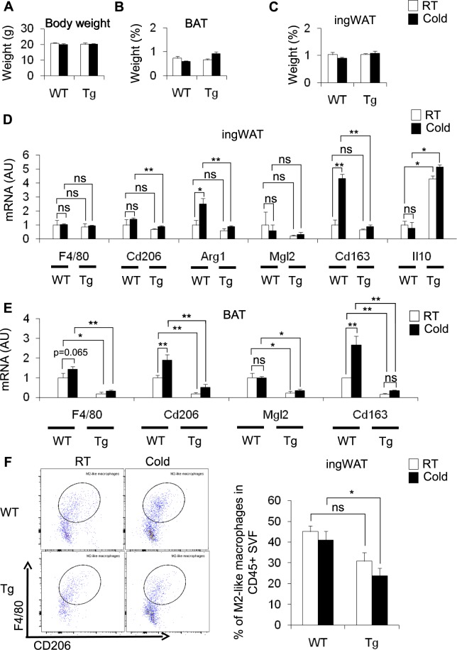
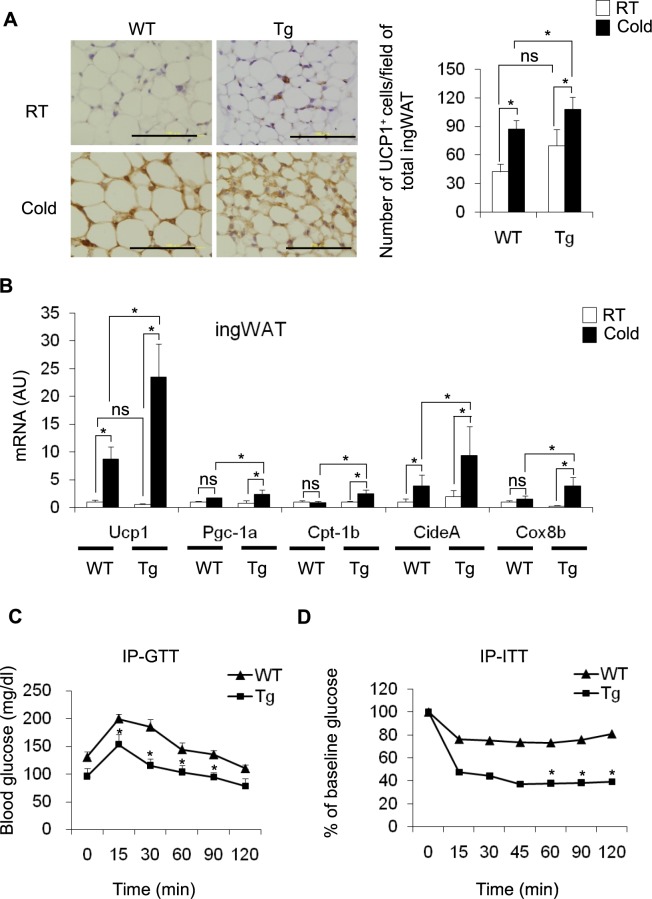
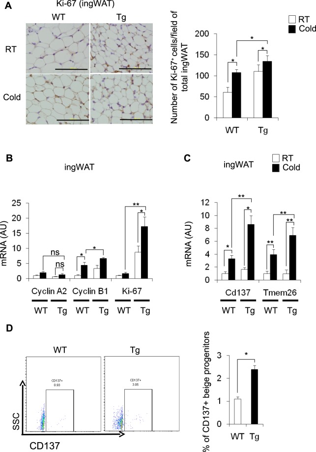
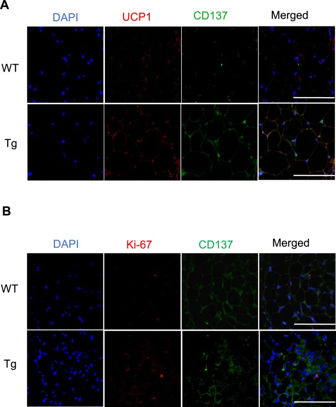
Beige adipocytes are an inducible form of thermogenic adipocytes that become interspersed within white adipose tissue (WAT) depots in response to cold exposure. Previous studies have shown that type 2 cytokines and M2 macrophages induce cold-induced browning in inguinal WAT (ingWAT) by producing catecholamines. Exactly how the conditional and partial depletion of CD206+ M2-like macrophages regulates the cold-induced browning of ingWAT, however, remains unknown. We examined the role of CD206+ M2-like macrophages in the cold-induced browning of WAT using genetically engineered CD206DTR mice, in which CD206+ M2-like macrophages were conditionally depleted. The partial depletion of CD206+ M2-like enhanced UCP1 expression in ingWAT, as shown by immunostaining, and also upregulated the expression of Ucp1 and other browning-related marker genes in ingWAT after cold exposure. A flow cytometry analysis showed that the partial depletion of CD206+ M2-like macrophages caused an increase in the number of beige progenitors in ingWAT in response to cold. Thus, we concluded that CD206+ M2-like macrophages inhibit the proliferation of beige progenitors and that the partial depletion of CD206+ M2-like macrophages releases this inhibition, thereby enhancing browning and insulin sensitivity.


Conflict of interest statement

The authors declare no competing interests.

Figures

Figure 1
Figure 1
Effect of cold stimulation on BAT and ingWAT in WT mice and DT-treated CD206DTR transgenic (Tg) mice. (AC) Changes in body weight (g) and adipose tissue weight (BAT and ingWAT) of WT and Tg mice at RT (white) and at a cold temperature (black) as the percentage of body weight (n = 4–6 mice per group). (D,E) mRNA expression of M2-like macrophage marker genes in the ingWAT and BAT of cold-stimulated WT and Tg mice compared with their littermate controls at RT (n = 3–5 mice per group). (F) Representative flow cytometric images of ingWAT from WT and Tg mice (RT vs. cold) (left panel) showing M2-like macrophages. The M2-like macrophages in CD45+ SVF are quantified in the right panel. After debris removal, live cells were gated for CD45+ cells (hematopoietic) and then for F4/80+CD206+ cells (M2-like macrophages) (n = 3–4 mice per group). An isotype control antibody was used as a negative control. The data are shown as the mean ± SEM. *p < 0.05, **p < 0.01, compared with their littermates, as determined using Student’s t-test (AU: arbitrary unit, ns: non-significant).
Figure 2
Figure 2
Partial depletion of CD206+ M2-like macrophages increases browning of ingWAT. (A) Representative images of paraffin sections of ingWAT from DT-treated WT and Tg mice after cold exposure (96 h) stained with anti-UCP1 antibody (left panel). UCP1+ cells were counted in 5 randomly selected microscopic fields (20×) and were expressed as the average cell number per microscopic field/total ingWAT weight (right panel) (n = 4–6 mice per group; scale bar, 100 μm). (B) mRNA expression of Ucp1 and other browning marker genes in the ingWAT of cold stimulated DT-treated WT and Tg mice (n = 4–6 mice per group). (C,D) Glucose concentrations from an intraperitoneal glucose tolerance test (IP-GTT) (n = 7 mice per group) and glucose concentrations from an intraperitoneal insulin tolerance test (IP-ITT) as the percent of the basal glucose level (n = 7–9 mice per group) in DT-treated WT and Tg mice maintained at a cold temperature. The data are shown as the mean ± SEM. *p < 0.05, **p < 0.01, compared with littermates, as determined using Student’s t-test.
Figure 3
Figure 3
Effect of cold exposure on cell cycle-related genes and beige progenitor genes in ingWAT of cold-stimulated Tg mice. (A) Representative images of paraffin sections of ingWAT of DT-treated WT and Tg mice after cold exposure stained with anti-Ki-67 antibody (left panel) and quantification of Ki-67+-cells/field for total ingWAT weight (right panel) (n = 4–6 mice per group; scale bar, 100 μm). (B,C) mRNA expression of cell cycle-related genes and beige progenitor marker genes in the ingWAT of cold-stimulated WT and Tg mice compared with their littermate controls at RT (n = 4–6 mice per group). (D) Representative flow cytometry images of ingWAT from WT and Tg mice (left panel). Quantification is shown in the right panel. After debris removal, live cells were gated for CD31/CD45 cells (lineage negative). Then, the lineage negative cells were further gated for the CD137+ population (n = 4–5 mice per group). An isotype control antibody was used as a negative control. The data are shown as the mean ± SEM. *p < 0.05, **p < 0.01, compared with littermates, as determined using Student’s t-test.
Figure 4
Figure 4
Immunofluorescence analysis of ingWAT. (A,B) Representative confocal images of paraffin sections of ingWAT from DT-treated WT and Tg mice after cold exposure stained with anti-UCP1 and anti-CD137 antibodies (A) and anti-CD137 & anti-Ki-67 antibodies. (B) The pictures were taken using a Leica TCS-SP-5, 40× (scale bar, 50 μm).

References

    1. Nedergaard J, Cannon B. The browning of white adipose tissue: some burning issues. Cell Metab. 2014;20:396–407. doi: 10.1016/j.cmet.2014.07.005. - DOI - PubMed
    1. Ikeda K, Maretich P, Kajimura S. The Common and Distinct Features of Brown and Beige Adipocytes. Trends Endocrinol Metab. 2018;29:191–200. doi: 10.1016/j.tem.2018.01.001. - DOI - PMC - PubMed
    1. Wynn TA, Chawla A, Pollard JW. Macrophage biology in development, homeostasis and disease. Nature. 2013;496:445–455. doi: 10.1038/nature12034. - DOI - PMC - PubMed
    1. Reilly SM, Saltiel AR. Adapting to obesity with adipose tissue inflammation. Nat Rev Endocrinol. 2017;13:633–643. doi: 10.1038/nrendo.2017.90. - DOI - PubMed
    1. Crewe C, An YA, Scherer PE. The ominous triad of adipose tissue dysfunction: inflammation, fibrosis, and impaired angiogenesis. J Clin Invest. 2017;127:74–82. doi: 10.1172/jci88883. - DOI - PMC - PubMed

Publication types