. 2018 Oct 1;9(1):4015.
doi: 10.1038/s41467-018-06307-w.

Mechanotransduction is required for establishing and maintaining mature inner hair cells and regulating efferent innervation

Affiliations

Mechanotransduction is required for establishing and maintaining mature inner hair cells and regulating efferent innervation

Laura F Corns et al. Nat Commun. .

Abstract

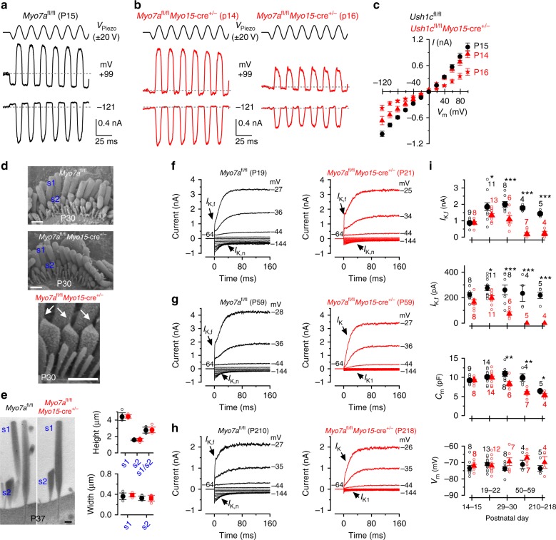
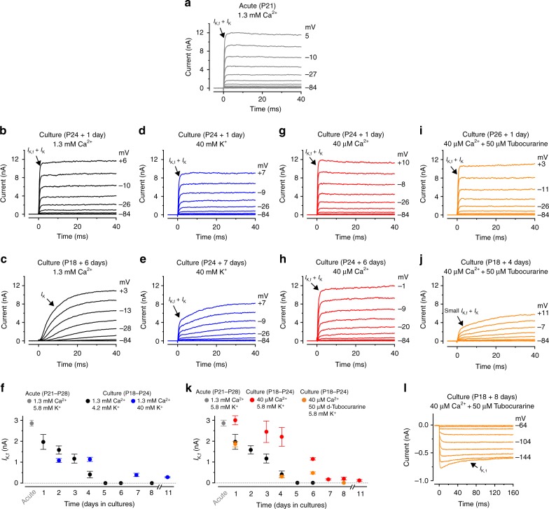
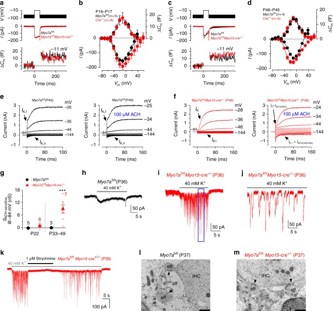
In the adult auditory organ, mechanoelectrical transducer (MET) channels are essential for transducing acoustic stimuli into electrical signals. In the absence of incoming sound, a fraction of the MET channels on top of the sensory hair cells are open, resulting in a sustained depolarizing current. By genetically manipulating the in vivo expression of molecular components of the MET apparatus, we show that during pre-hearing stages the MET current is essential for establishing the electrophysiological properties of mature inner hair cells (IHCs). If the MET current is abolished in adult IHCs, they revert into cells showing electrical and morphological features characteristic of pre-hearing IHCs, including the re-establishment of cholinergic efferent innervation. The MET current is thus critical for the maintenance of the functional properties of adult IHCs, implying a degree of plasticity in the mature auditory system in response to the absence of normal transduction of acoustic signals.


Conflict of interest statement

The authors declare no competing interests.

Figures

Fig. 1
Fig. 1
Current and voltage responses from IHCs of Pcdh15 mutant mice. a, b Potassium currents recorded from IHCs of control Pcdh15+/av3j (a) and littermate mutant Pcdh15av3j/av3j mice (b) using 10 mV depolarizing voltage steps from –84 mV to the various test potentials shown by some of the traces. The adult-type currents (IK,f and IK,n) were only present in IHCs from control Pcdh15+/av3j mice (a). IHCs from Pcdh15av3j/av3j mice retained the currents characteristic of immature cells (b, IK and IK1). The absence of the rapidly activating IK,f in Pcdh15 av3j/av3j IHCs is also evident when comparing the activation time course of the total outward currents on an expanded time scale (see insets). a, b are representative recordings from 10 (3 mice) and 12 (3 mice) IHCs, respectively. c, d Voltage responses elicited by applying depolarizing current injections to control, Pcdh15+/av3j c and mutant, Pcdh15av3j/av3j d IHCs from their resting membrane potentials. Depolarizing current injections caused slow APs in the Pcdh15avj3/av3j mutant IHC. c, d are representative recordings from 8 (3 mice) and 15 (3 mice) IHCs, respectively. Recordings were performed near body temperature
Fig. 2
Fig. 2
Disappearance of the MET current in IHCs of Ush1cfl/fl conditional KO mice. a MET currents recorded in P7 IHCs from control Ush1cfl/fl and knockout Ush1cfl/flMyo15-cre+/- mice by displacing the hair bundles while applying 20 mV voltage steps between −121 mV to + 99 mV. For clarity, only the traces at −121 mV and + 99 mV are shown (dashed lines: holding current; VPiezo: driver voltage to the fluid jet). b Maximum MET current-voltage curves obtained from P7 (left) and P8 (right) Ush1cfl/fl and Ush1cfl/flMyo15-cre+/- IHCs (3 and 6 mice, respectively). c MET currents in P10 IHCs from both genotypes. Protocol as in a. d Maximum MET currents obtained from 9 Ush1cfl/fl and 17 Ush1cfl/flMyo15-cre+/- (P10-P12) IHCs (4 and 8 mice, respectively). e SEM images of P15 IHC bundle from both genotypes. Scale bars, 1 μm. f TEM images (left) of P25 IHC stereocilia from both genotypes. Scale bars, 400 nm. Right panels show the height and width of the tallest (s1) and second tallest (s2) stereocilia in P25 IHCs from Ush1cfl/fl and Ush1cfl/flMyo15-cre+/- mice. g Depolarizing current used to elicit repetitive APs in Ush1cfl/fl (P9-P10, 8 mice) and Ush1cfl/flMyo15-cre+/- (P9-P11, 7 mice) IHCs. Experimental conditions: body temperature (BT: 34–37 °C); 1.3 mM extracellular Ca2+. h Size of the resting MET current (IMET) recorded from Ush1cfl/fl IHCs (P9-P11, 2 mice) and Ush1cfl/flMyo15-cre+/- (P10-P11, 5 mice) IHCs. Room temperature (RT: 22–24 °C, left bars); 1.3 mM extracellular Ca2+. IMET was interpolated from its current-voltage relation near the Vm of IHCs (−73 mV). Right bars show the RT values corrected for BT using a Q10 of 1.45 (ref. ). i Size of the resting IMET at −73 mV from Ush1cfl/fl (P10, 3 mice) and Ush1cfl/flMyo15-cre+/- (P10, 3 mice) IHCs using the endolymph-like Ca2+ concentration (0.3 mM: see panel j). Left and right bars are as described in h. j IMET recorded at −81 mV at P10 from Ush1cfl/fl and Ush1cfl/flMyo15-cre+/- IHCs in 0.3 mM Ca2+. Protocol as in a. Number of IHCs in gi is shown above the columns. Average data is shown as mean ± SEM in b, d, g, h or mean ± SD in f
Fig. 3
Fig. 3
IHCs from Ush1cfl/flMyo15-cre+/- conditional knockout mice fail to mature. a, b Potassium currents recorded from P15 (upper panels) and P29 (lower panels) IHCs from control Ush1cfl/fl (a) and Ush1cfl/flMyo15-cre+/- (b) mice using 10 mV depolarizing and hyperpolarizing voltage steps from −64 mV. Note that the K+ currents characteristic of adult IHCs (IK,f and IK,n) were only present in IHCs from control Ush1cfl/fl mice (a). IHCs from Ush1cfl/flMyo15-cre+/- mice retained the K+ currents characteristic of pre-hearing stages (IK and IK1). c, d Voltage responses elicited by applying hyperpolarizing and depolarizing current injections from the respective membrane potentials in P15 IHCs of control Ush1cfl/fl (c) and Ush1cfl/flMyo15-cre+/- (d) mice. Note that APs are still present in post-hearing Ush1cfl/flMyo15-cre+/- IHCs. For more details see Supplementary Table 2. e, f Mean ABR thresholds ( ±SD ) for clicks and pure tone stimulation from 3 kHz to 24 kHz obtained from control Ush1cfl/fl and littermate Ush1cfl/flMyo15-cre+/- mice at P16 (e) and P37-45 (f). Ush1cfl/flMyo15-cre+/- mice are almost completely deaf already at P16. Open symbols represent data from single mice. Closed symbols represent averages (mean ± SEM)
Fig. 4
Fig. 4
IHCs from Myo7afl/flMyo15-cre+/- conditional knockout mice acquire their mature phenotype. a, b MET currents from IHCs of control Myo7afl/fl (left, P10) and knockout Myo7afl/flMyo15-cre+/- (right, P9) mice. The experimental protocol is as in Fig. 2. c Cochlear whole mount preparations from P10 Myo7afl/fl (control, upper panel) and Myo7afl/flMyo15-cre+/- (lower panel) mice, immunostained for Myo7a (green) and for F-actin with phalloidin (red). Myo7a is detected in both OHCs and IHCs in the Myo7afl/fl mouse, but not in the Myo7afl/flMyo15-cre+/- mouse (but see also Supplementary Fig. 4 for one example of normal hair cell Myo7a labeling in a P10 Myo7afl/flMyo15-cre+/- mouse). Scale bars, 5 µm. d IHC voltage response recorded during the superfusion of 0.3 mM Ca2+ alone and together with the MET channel blocker DHS (0.2 mM) in a Myo7afl/flMyo15-cre+/- mouse. The MET depolarizing current substantially contributes to the IHC membrane potential. e Images showing relative fluo-4 fluorescence changes (ΔF/F0) before (left) during (middle) and after (right) application of an extracellular solution containing 0.3 mM Ca2+. Images were obtained as maximal back projection (200 frames, 6.6 s). Scale bar, 15 µm. f Representative ΔF/F0 traces from two IHCs (from images in panel e) during bath application of 0.3 mM Ca2+. Traces are computed as pixel averages of regions of interest centered on IHCs. Ca2+ spikes are evident in IHCs; however, the relative long fluorescence decay time constant of the Ca2+ signal, prevents the isolation of single APs during high-frequency bursting. g, h Current responses in post-hearing IHCs (P15) from control (g) and Myo7afl/flMyo15-cre+/- (h) mice. Note that both IK,f and IK,n are present in IHCs from both genotypes. Voltage protocol as in Fig. 3a. i Voltage responses recorded from P15 IHCs of control Myo7afl/fl (top panel) and Myo7afl/flMyo15-cre+/- (lower panel) P15 IHCs (protocol as in Fig. 3c). Panel (g, j: Myo7afl/fl) and (b, j: Myo7afl/flMyo15-cre+/-) are representative recordings from 9 IHCs (4 mice) and 8 IHCs (4 mice), respectively. j Mean ABR thresholds ( ± SD) for clicks and frequency-specific pure tone stimulation from 3 Hz to 24 kHz obtained from control Myo7afl/fl and littermate Myo7afl/flMyo15-cre+/- mice at P15
Fig. 5
Fig. 5
The functional MET current is required for retaining mature IHC characteristics. a, b MET currents recorded from mature IHCs of control Myo7afl/fl (a, P15) and Myo7afl/flMyo15-cre+/- (b, P14 and P16) mice. Protocol as in Fig. 2a. c Peak-to-peak MET current-voltage curves obtained from six P15 Myo7afl/fl (2 mice), four P14 Myo7afl/flMyo15-cre+/- and six P16 Myo7afl/flMyo15-cre+/- IHCs (3 mice). d Scanning electron microscopy images showing the stereociliary bundle of apical-coil IHC from P30 control Myo7afl/fl and Myo7afl/flMyo15-cre+/- mice. s1 and s2 represent the tallest and the second tallest stereocilia, respectively. The arrows in the middle panel highlights that tip links, which are required to gate the MET current, are present in Myo7afl/flMyo15-cre+/- IHCs. Scale bars, 1 μm. e Transmission electron microscopy images (left) showing the stereocilia s1 and s2 (see panel d) of apical-coil IHCs from P37 control Myo7afl/fl and Myo7afl/flMyo15-cre+/- mice. Scale bars, 400 nm. The right panels show that the height and width of s1 and s2 stereocilia in IHCs are comparable between the two genotypes (Myo7afl/fl s1: height 4.42 ± 0.48 μm, width 0.36 ± 0.07 μm, n = 6 IHCs, s2: height 1.58 ± 0.10 μm, width 0.32 ± 0.04 μm, n = 7, s1/s2: 2.77 ± 0.36, n = 6; Myo7afl/flMyo15-cre+/- s1: 4.44 ± 0.20 μm, P < 0.8915, 0.38 ± 0.03 μm, P < 0.4441, n = 7, s2: 1.61 ± 0.18 μm, P < 0.7849, 0.33 ± 0.06 μm, P < 0.8959, n = 7, s1/s2: 2.80 ± 0.35, P < 0.9059, n = 6). fh Potassium currents recorded from IHCs of young adult (f, P19 and P21), adult (g, P59) and aged (h, P210 and P218) control Myo7afl/fl (left panels) and Myo7afl/flMyo15-cre+/- (right panels) mice. Protocols are as in Fig. 3a. i Size of IK,f, IK,n, membrane capacitance (Cm) and resting membrane potential (Vm) as a function of postnatal age. Number of IHC investigated is shown above or below the symbols (for more details see Supplementary Table 3). Statistical tests are performed with two-way ANOVA with Bonferroni correction. Average data is shown as mean ± SEM (c, i) or mean ± SD (e)
Fig. 6
Fig. 6
The resting MET current drives the maintenance of mature IHC K+ currents in cochlear cultures. a Potassium outward currents recorded from a P21 IHC of an acutely dissected gerbil cochlea. Recordings were performed using a perilymph-like solution (1.3 mM extracellular Ca2+; 5.8 mM K+). Currents were elicited using 10 mV depolarizing voltage steps from −84 mV. Note the fast activating IK,f characteristic of adult IHCs (arrow). b, c Currents recorded from IHCs maintained in cochlear culture for 1 day (b) or 6 days (c). The culture medium bathing the cochlea (see Methods) contained 1.3 mM Ca2+ and 4.2 mM K+. d, e Currents recorded from IHCs maintained in culture for 2 day (d) or 7 days (e). Culture medium contained 1.3 mM Ca2+ and 40 mM K+. f Size of IK,f recorded from IHCs of acutely isolated gerbil cochleae (gray circle, n = 6, P21-P28, 3 gerbils) or maintained in culture. Culture conditions were: (1) normal medium (black circles, number of IHCs from left to right: 7,3,4,10,2,6,7; from 9 cochlear cultures; gerbil age at the day of culturing: P18-P24); 2) medium supplemented to 40 mM K+ (blue circles, number of IHCs: 6,6,5,7, from 4 cochlear cultures, P18-P24). g, h Currents recorded from IHCs maintained in culture for 1 day (g) or 6 days (h) with a medium containing 40 µM Ca2+. i, j, l Currents from IHCs maintained in cochlear culture for 1 day (i), 4 days (j) or 8 days (l) with a medium containing 40 µM Ca2+ and 50 μM of the MET channel blocker d-tubocurarine. The inward rectifier K+ current (IK1) l was obtained using 10 mV hyperpolarizing voltage steps from −64 mV. k Size of IK,f recorded from IHCs of acutely dissected cochleae (gray circle: same as in panel f), maintained in culture with a normal medium (black circles: as in panel f), a medium containing 40 µM Ca2+ (red circles: number of IHCs: 5,10,9,5,3,5,8, from 9 cochlear cultures, P18-P24) and a medium containing 40 µM Ca2+ and 50 μM d-tubocurarine (orange circles: 4,6,6,4, from 4 cochlear cultures, P18-P24). Average data is shown as mean ± SEM
Fig. 7
Fig. 7
IHC exocytosis is unaffected in adult Myo7afl/flMyo15-cre+/- mice but efferent input is altered. ad Calcium current (ICa) and corresponding changes in membrane capacitance (ΔCm) recorded from IHCs of P16-P17 (a, b) and P48-P49 (c, d) control Myo7afl/fl (black) and knockout Myo7afl/flMyo15-cre+/- mice (red). Recordings were obtained in response to 50 ms voltage steps (10 mV increments) from −81 mV and using 1.3 mM extracellular Ca2+ and at body temperature. For clarity, only maximal responses at −11 mV are shown. Panels b and d shows average peak ICa (bottom) and ΔCm (top) curve from control Myo7afl/fl and Myo7afl/flMyo15-cre+/- IHCs. e, f Membrane currents recorded from adult IHCs of control Myo7afl/fl (e) and Myo7afl/flMyo15-cre+/- (f) mice before (left panels) and during superfusion of ACh (right panels). ACh elicited an instantaneous current only in the Myo7afl/flMyo15-cre+/- IHC. g Steady-state slope conductance of the ACh-sensitive current (gACh-sensitive) at different age ranges in control Myo7afl/fl (black symbols) and Myo7afl/fl Myo15-cre+/- (red symbols) IHCs. The isolated gACh-sensitive was obtained by subtracting the control current (left panels in e, f) from that in the presence of 100 μM ACh (right panels). Number of IHCs is shown near the symbols (2 Myo7afl/fl mice at P22 and P33-P49; 3 Myo7afl/flMyo15-cre+/- mice at P22; 5 Myo7afl/flMyo15-cre+/-mice at P34-P48). hk Voltage-clamp recordings obtained from adult IHCs of control Myo7afl/fl (h, P36) and Myo7afl/flMyo15-cre+/- (i, j, P36; k P39) mice during the superfusion of 40 mM extracellular K+. Synaptic currents were only evoked in Myo7afl/flMyo15-cre+/- IHCs. Panel j shows an expanded time scale of the blue area shown in panel (i). Panel k shows the effect of 1 μM strychnine on the ACh-induced synaptic currents. l, m TEM showing the IHC synaptic region from P37 control Myo7afl/fl (l) and Myo7afl/flMyo15-cre+/- (m) mice. IHCs from Myo7afl/fl mice showed the characteristic synaptic organization of mature cells with efferent terminals forming axo-dendritic contact with the afferent fibers, as seen by the presence of active zones (l, black arrows). Scale bars, 1 μm. In Myo7afl/flMyo15-cre+/- mice efferent fibers make direct axo-somatic contact with the IHC (m), which is characteristic of pre-hearing IHCs. Average data is shown as mean ± SEM

References

    1. Goodyear RJ, Marcotti W, Kros CJ, Richardson GP. Development and properties of stereociliary link types in hair cells of the mouse cochlea. J. Comp. Neurol. 2005;485:75–85. doi: 10.1002/cne.20513. - DOI - PubMed
    1. Kazmierczak P, et al. Cadherin 23 and protocadherin 15 interact to form tip-link filaments in sensory hair cells. Nature. 2007;449:87–91. doi: 10.1038/nature06091. - DOI - PubMed
    1. Barr-Gillespie PG. Assembly of hair bundles, an amazing problem for cell biology. Mol. Biol. Cell. 2015;26:2727–2732. doi: 10.1091/mbc.e14-04-0940. - DOI - PMC - PubMed
    1. Corns LF, Johnson SL, Kros CJ, Marcotti W. Calcium entry into stereocilia drives adaptation of the mechanoelectrical transducer current of mammalian cochlear hair cells. Proc. Natl Acad. Sci. USA. 2014;111:14918–14923. doi: 10.1073/pnas.1409920111. - DOI - PMC - PubMed
    1. Johnson SL, Kennedy HJ, Holley MC, Fettiplace R, Marcotti W. The resting transducer current drives spontaneous activity in prehearing mammalian cochlear inner hair cells. J. Neurosci. 2012;32:10479–10483. doi: 10.1523/JNEUROSCI.0803-12.2012. - DOI - PMC - PubMed

Publication types

MeSH terms

Substances