. 2018 Oct;25(10):951-957.
doi: 10.1038/s41594-018-0131-8. Epub 2018 Oct 1.

DNA G-quadruplex structures mold the DNA methylome

Affiliations

DNA G-quadruplex structures mold the DNA methylome

Shi-Qing Mao et al. Nat Struct Mol Biol. 2018 Oct.

Abstract

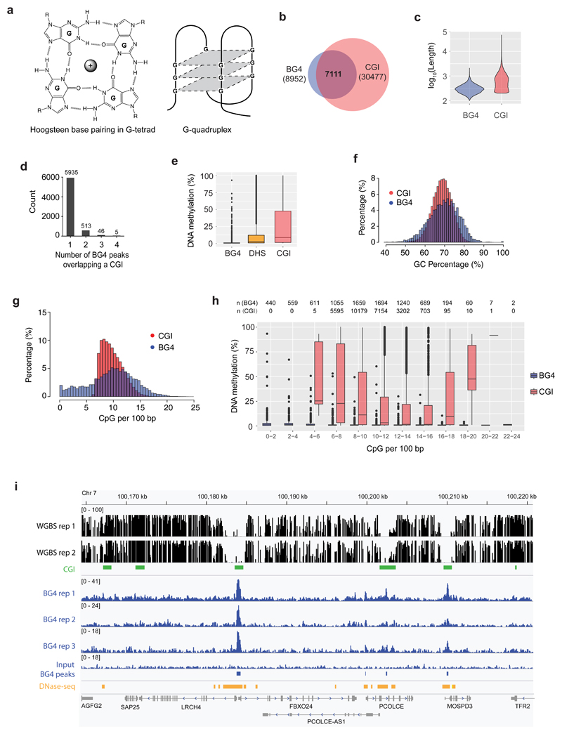
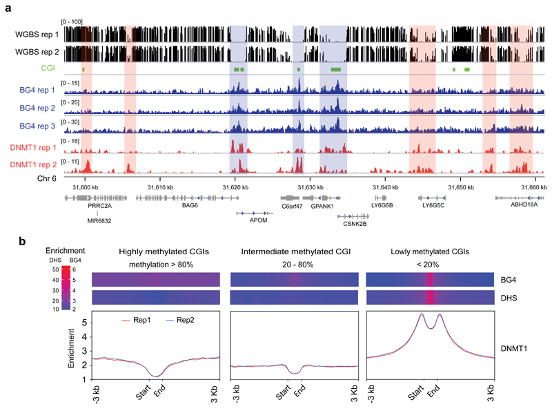
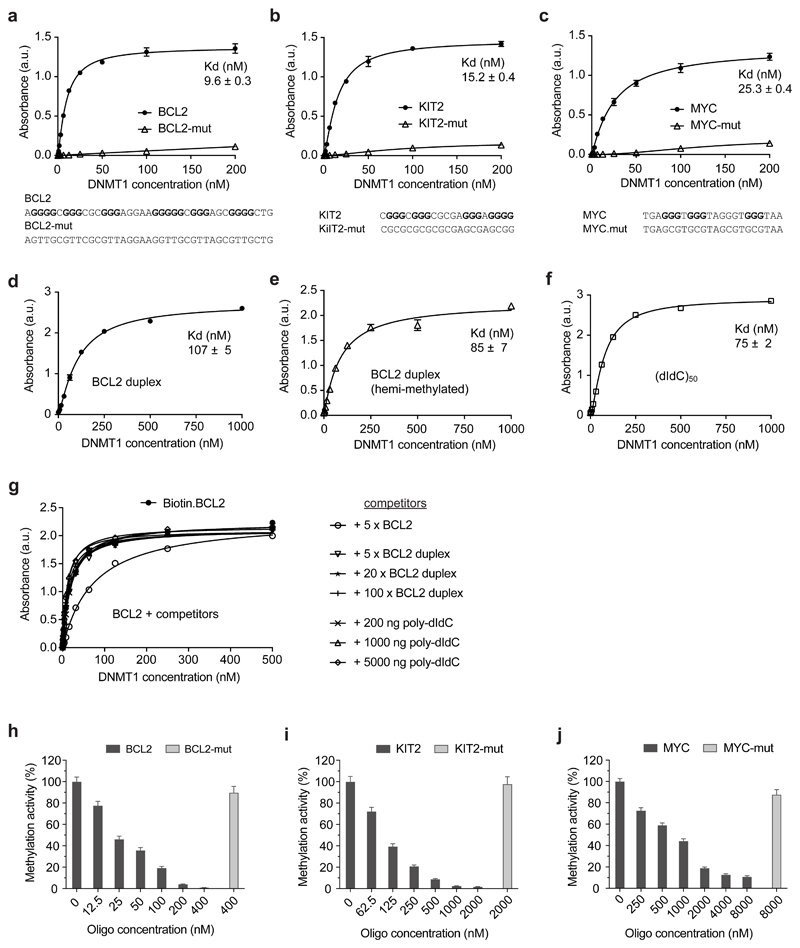
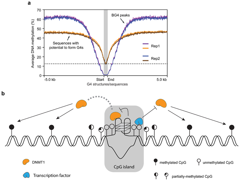
Control of DNA methylation level is critical for gene regulation, and the factors that govern hypomethylation at CpG islands (CGIs) are still being uncovered. Here, we provide evidence that G-quadruplex (G4) DNA secondary structures are genomic features that influence methylation at CGIs. We show that the presence of G4 structure is tightly associated with CGI hypomethylation in the human genome. Surprisingly, we find that these G4 sites are enriched for DNA methyltransferase 1 (DNMT1) occupancy, which is consistent with our biophysical observations that DNMT1 exhibits higher binding affinity for G4s as compared to duplex, hemi-methylated, or single-stranded DNA. The biochemical assays also show that the G4 structure itself, rather than sequence, inhibits DNMT1 enzymatic activity. Based on these data, we propose that G4 formation sequesters DNMT1 thereby protecting certain CGIs from methylation and inhibiting local methylation.


Conflict of interest statement

Competing interests

SB is an advisor and shareholder of Cambridge Epigenetix limited.

Figures

Figure 1
Figure 1. G4 formation is associated with hypomethylation at CGIs
a) A G-tetrad stabilized by Hoogsteen hydrogen bonding and a central monovalent cation (left). Schematic representations of a three-tetrad G4 structure (Right). b) Venn diagram illustrating the overlap of G4 structure formation (BG4 peaks) and CGIs. c) Violin plot showing size distribution of BG4 peaks and CGIs. d) Count of BG4 peaks overlapping a CGI. e) Box and whisker plot showing the average methylation for BG4 peaks (n = 8,210), DHSs (n = 142,115) and CGIs (n = 27,073). Centre line represents the median value separating upper and lower quartiles in the box, whiskers indicate 1.5× interquartile range (IQR), points are actual values of outliers. Note that methylation level at CpG sites with less than 5x coverage is considered unreliable and discarded. f) Histogram showing the distribution of BG4 peaks and CGIs relative to percentage of GC. g) Histogram showing the distribution of BG4 peaks and CGIs relative to percentage of CpGs per 100 bp. h) Box and whisker plot showing the methylation levels for BG4 peaks and CGIs at different CpG densities. Note that by definition there are no CGIs at a CpG density < 5 CpGs/100bp and that at > 20 CpGs/100bp there are few CGIs (1) and BG4 (36) peaks to consider. The number of CGI regions and BG4 peaks in each category are presented on top of the plot. i) An IGV screen shot illustrating the co-incidence of BG4 peaks (blue) with hypomethylated promoter CGIs (green) and DHSs (orange) for a representative genome region from Chr 7. Shown are normalised signal. Whole genome bisulfite sequence tracks are in black (top). RefGene tracks are in grey (bottom).
Figure 2
Figure 2. DNMT1 is recruited to BG4 peaks associated with low methylation
a) An IGV screen shot showing the co-incidence (blue-masked) of BG4 peaks (blue) with DNMT1 ChIP-seq peaks (red) and CGIs (green) at hypomethylated region from Chr 6. Orange-masked regions are hypermethylated and enriched with DNMT1 presence, but not BG4 signal. Whole genome bisulfite sequence tracks in black (top). b) Binding profile of DNMT1 shown across CGIs with low (less than 20%, n = 16,523), intermediate (between 20% and 80%, n = 6,042) and high (more than 80%, n = 4,266) methylation. Y-axis shows the number of reads in the ChIP normalised to 1 of sequencing depth (also known as Reads Per Genomic Content (RPGC), more details in computational methods). Replicate 1 and 2 are indicated in red and blue respectively. Above each plot is a heat map showing the enrichment of BG4 peaks and DHSs across the respective regions. The heat maps show RPGC of active chromatin marks (DHSs) and BG4 peaks on these three classes of CGIs.
Figure 3
Figure 3. DNMT1 selectively binds and is inhibited by G4 structures
a-f) ELISA assays testing the binding of recombinant DNMT1 to G4 structure and control oligonucleotides. Binding curves for: a) BCL2 G4 and non-G4-forming control (BCL2-mut); b) KIT2 G4 and non-G4-forming control (KIT2-mut); c) MYC G4 and non-G4-forming control (MYC-mut); d) BCL2 duplex DNA; e) BCL2 hemi-methylated duplex DNA; f) poly(dI-dC), 100 nt. Absorbance was measured at 450 nm. a.u., arbitrary unit. Sequences of oligonucleotides are given below the graphs. g) Binding curve of BCL2 G4 in presence of different concentration of BCL2 duplex or poly(dIdC)n. h-j) Relative methylation activity of recombinant DNMT1 in presence of G4 structure and control oligonucleotides: h) BCL2 G4 and BCL2-mut; i) KIT2 G4 and KIT2-mut; j) MYC G4 and MYC-mut. Shown are mean ± s.d., n = 3 independent experiments in all plots but g (n = 2).
Figure 4
Figure 4. Recruitment of DNMT1 by G4 structures shapes the methylome in G-rich regions
a) Plot showing the average methylation profile centred around G4 forming regions (red and blue are replicates 1 and 2 respectively, n = 7,491) or G4 sequences without structure (orange and green are replicates 1 and 2 respectively, n = 36,015). The plot extends ± 5 Kb from the centre. The dotted line denotes the lowest methylation level of G4 sequence without structure. b) Proposed model for potential involvement of G4 structures and methylation control at CGIs: i) G4 structures sequester DNMT1 due to high affinity binding; ii) G4 structures inhibit the methylation activity of DNMT1. Together with the binding of transcription factors, G4 structures contribute to protection of CGIs from methylation.

References

    1. Smith ZD, Meissner A. DNA methylation: roles in mammalian development. Nat Rev Genet. 2013;14:204–20. - PubMed
    1. Feinberg AP, Tycko B. The history of cancer epigenetics. Nat Rev Cancer. 2004;4 - PubMed
    1. Li E, Bestor TH, Jaenisch R. Targeted mutation of the DNA methyltransferase gene results in embryonic lethality. Cell. 1992;69:915–926. - PubMed
    1. Okano M, Bell DW, Haber DA, Li E. DNA methyltransferases Dnmt3a and Dnmt3b are essential for de novo methylation and mammalian development. Cell. 1999;99:247–257. - PubMed
    1. Liao J, et al. Targeted disruption of DNMT1, DNMT3A and DNMT3B in human embryonic stem cells. Nat Genet. 2015;47:469–478. - PMC - PubMed

Publication types