

The Ethics of Interstitial and Cesarean Scar Ectopic Pregnancies: Four Case Studies and a Review of the Literature
- PMID: 30275610
- PMCID: PMC6161235
- DOI: 10.1177/0024363918788858
The Ethics of Interstitial and Cesarean Scar Ectopic Pregnancies: Four Case Studies and a Review of the Literature
Abstract
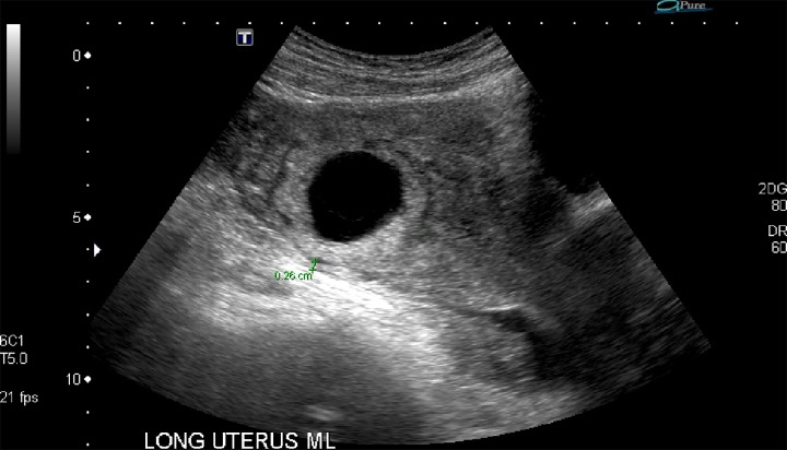
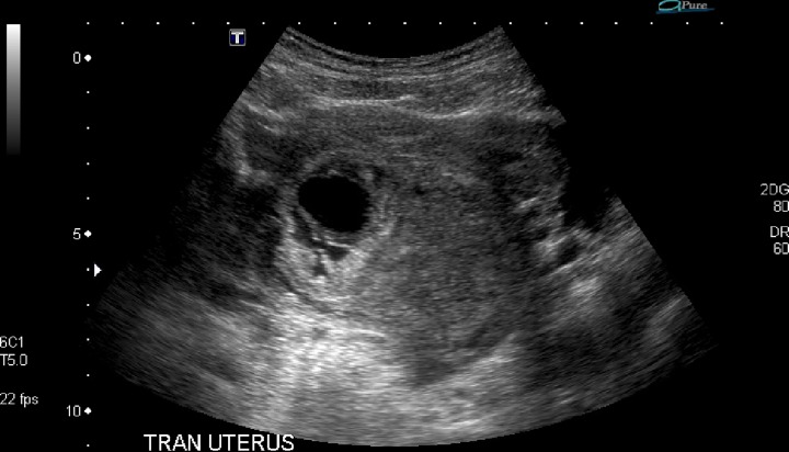
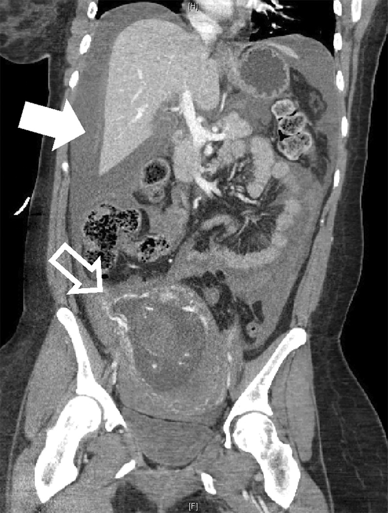
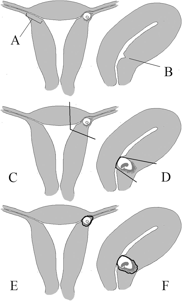
Catholic bioethicists have extensively addressed extrauterine tubal pregnancies, which represent the great majority of ectopic pregnancies. However, additional management options have been developed for the other 7-10 percent of ectopic pregnancies. Using two cases of interstitial pregnancy and two cases of cesarean scar pregnancy (CSP) seen at a Catholic tertiary care center, this article discusses options including expectant management, systemic methotrexate, intragestational methotrexate, intragestational potassium chloride, uterine artery embolization, dilation and curettage (D&C), vasopressin use, cornuostomy, cornual wedge resection, CSP evacuation, CSP scar excision, CSP salvage, and hysterectomy. Cornual wedge resection, vasopressin use, and CSP scar excision are morally acceptable; less clearly licit are aspiration of gestational sac contents, cornuostomy, gestational excision for CSPs, and methotrexate. Certainly illicit are any techniques leading to direct abortion such as D&Cs on live embryos or fetuses, double-balloon catheter placement, and use of potassium chloride. Summary: An ectopic pregnancy is any pregnancy outside the uterus. These are dangerous because the pregnancy can burst out of its abnormal location and cause life-threatening internal bleeding. Most are in the part of the fallopian tube outside the uterus, but there are other types, including interstitial pregnancies (located in the part of the tube tunneling through the uterine wall) and cesarean scar pregnancies (buried in the uterine scar where the cut for a C-section was made). This article lists the ways that physicians prevent women from dying from interstitial and cesarean scar pregnancies and proposes which treatments are morally acceptable.
Keywords: Bioethics; Cesarean scar pregnancy; Ectopic pregnancy; Indirect abortion; Interstitial pregnancy; Methotrexate; Vasopressin.
Conflict of interest statement
Declaration of Conflicting Interests: The author(s) declared no potential conflicts of interest with respect to the research, authorship, and/or publication of this article.
Figures
Similar articles
-
Cesarean Scar Pregnancy, Incidence, and Recurrence: Five-Year Experience at a Single Tertiary Care Referral Center.Obstet Gynecol. 2018 Nov;132(5):1285-1295. doi: 10.1097/AOG.0000000000002940. Obstet Gynecol. 2018. PMID: 30303911
-
Exclusive use of intrasac potassium chloride and methotrexate for treating cesarean scar pregnancy: effectiveness and subsequent fecundity.Hum Reprod Open. 2020 May 18;2020(2):hoaa025. doi: 10.1093/hropen/hoaa025. eCollection 2020. Hum Reprod Open. 2020. PMID: 32685702 Free PMC article.
-
Cesarean scar ectopic pregnancies: etiology, diagnosis, and management.Obstet Gynecol. 2006 Jun;107(6):1373-81. doi: 10.1097/01.AOG.0000218690.24494.ce. Obstet Gynecol. 2006. PMID: 16738166
-
Different treatment modalities for cesarean scar pregnancies: a single-center experience and literature review.Arch Gynecol Obstet. 2021 May;303(5):1143-1151. doi: 10.1007/s00404-020-05831-9. Epub 2020 Oct 13. Arch Gynecol Obstet. 2021. PMID: 33048187 Review.
-
Reproductive outcomes following cesarean scar pregnancy - a case series and review of the literature.Eur J Obstet Gynecol Reprod Biol. 2016 May;200:102-7. doi: 10.1016/j.ejogrb.2016.02.039. Epub 2016 Mar 8. Eur J Obstet Gynecol Reprod Biol. 2016. PMID: 27014853 Review.
Cited by
-
Cesarean section scar pregnancy: Challenges in choosing treatment approach.Clin Case Rep. 2021 Aug 16;9(8):e04592. doi: 10.1002/ccr3.4592. eCollection 2021 Aug. Clin Case Rep. 2021. PMID: 34429990 Free PMC article.
References
-
- Arleo Elizabeth Kagan, Defilippis Ersilia M. 2014. “Cornual, Interstitial, and Angular Pregnancies: Clarifying the Terms and a Review of the Literature.” Clinical Imaging 38, no. 6: 763–70. - PubMed
-
- Ashley Benedict M., O’Rourke Kevin D. 2002. Ethics of Health Care: An Introductory Textbook. Washington, DC: Georgetown UP.
-
- Aquinas Thomas. 1920. The “Summa Theologica” of St. Thomas Aquinas. London, UK: Burns Oates & Washbourne, Web. May 6, 2017.
-
- Bernstein Ira M. 2002. “Uterine Artery Hemodynamic Adaptations through the Menstrual Cycle into Early Pregnancy.” Obstetrics & Gynecology 99, no. 4: 620–24. - PubMed
LinkOut - more resources
Full Text Sources