Review
. 2018 Sep 26:22:26.
doi: 10.1186/s40824-018-0136-8. eCollection 2018.

Micro-CT - a digital 3D microstructural voyage into scaffolds: a systematic review of the reported methods and results

Affiliations
Review

Micro-CT - a digital 3D microstructural voyage into scaffolds: a systematic review of the reported methods and results

Ibrahim Fatih Cengiz et al. Biomater Res. .

Abstract

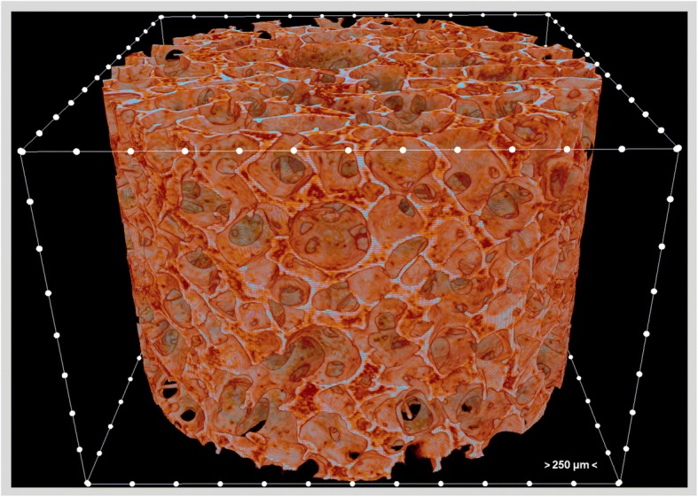
Background: Cell behavior is the key to tissue regeneration. Given the fact that most of the cells used in tissue engineering are anchorage-dependent, their behavior including adhesion, growth, migration, matrix synthesis, and differentiation is related to the design of the scaffolds. Thus, characterization of the scaffolds is highly required. Micro-computed tomography (micro-CT) provides a powerful platform to analyze, visualize, and explore any portion of interest in the scaffold in a 3D fashion without cutting or destroying it with the benefit of almost no sample preparation need.

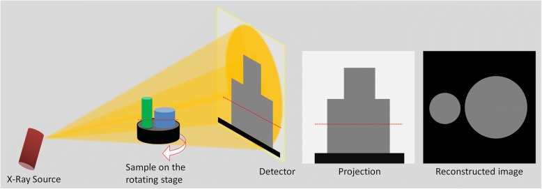
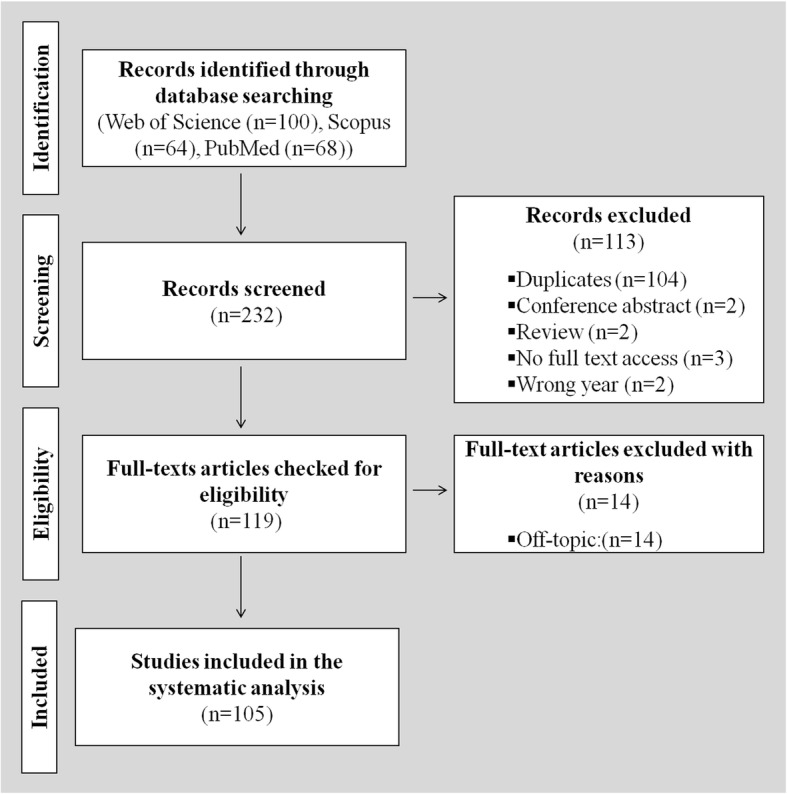
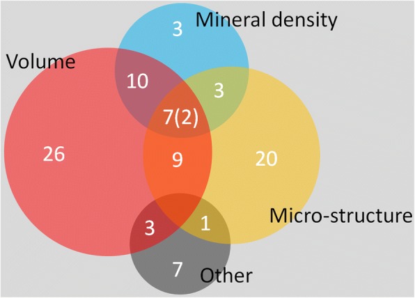
Main body: This review highlights the relationship between the scaffold microstructure and cell behavior, and provides the basics of the micro-CT method. In this work, we also analyzed the original papers that were published in 2016 through a systematic search to address the need for specific improvements in the methods section of the papers including the amount of provided information from the obtained results.

Conclusion: Micro-CT offers a unique microstructural analysis of biomaterials, notwithstanding the associated challenges and limitations. Future studies that will include micro-CT characterization of scaffolds should report the important details of the method, and the derived quantitative and qualitative information can be maximized.

Keywords: Microstructure; Mineral density; Scaffolds; Systematic review; Tissue engineering.


Conflict of interest statement

Not applicable.Not applicable.The authors declare that they have no competing interests.Springer Nature remains neutral with regard to jurisdictional claims in published maps and institutional affiliations.

Figures

Fig. 1
Fig. 1
A 3D micro-CT image of the polycaprolactone-polyurethane scaffold. The distance between two adjacent white dots is 250 μm
Fig. 2
Fig. 2
Schematic illustration showing the basics of micro-CT. Cone-beam X-rays travel from the source to the detector through the sample with attenuation, and a gray-scale projection image is acquired at each rotation angle. Projection images are then reconstructed, and the reconstructed image dataset is used for analysis. The red dashed line indicates the vertical position of the reconstructed image, i.e., the cross-sectional image
Fig. 3
Fig. 3
Binarization of reconstructed micro-CT images. Gray-scale reconstructed 2D image of a silk-based tissue engineering scaffold with a circular region of interest (ø 3 mm) (a), half-tone views (b, d, and f) and the corresponding binary images (c, e, and g) respectively if no gray-scale value is included that yields to complete black, if entire gray-scale values are included that yields to complete white, and if the right gray-scale values (that is in this case 38-255) are obtained by global thresholding that yields to a binary image showing the microstructure of the scaffold
Fig. 4
Fig. 4
The flowchart of the systematic search
Fig. 5
Fig. 5
Venn diagram showing the number of the papers that reported quantitative results regarding the volume, microstructure, mineral density and other measures. Two of the seven papers that reported volume, microstructure, and mineral density also reported additional results. The sizes of the circles in the diagrams are directly proportional to the number of the associated papers

References

    1. Wegst UG, Bai H, Saiz E, Tomsia AP, Ritchie RO. Bioinspired structural materials. Nat Mater. 2015;14(1):23–36. doi: 10.1038/nmat4089. - DOI - PubMed
    1. Lee J, Cuddihy MJ, Kotov NA. Three-dimensional cell culture matrices: State of the art. Tissue Eng Part B-Re. 2008;14(1):61–86. doi: 10.1089/teb.2007.0150. - DOI - PubMed
    1. Baker BM, Chen CS. Deconstructing the third dimension - How 3D culture microenvironments alter cellular cues. J Cell Sci. 2012;125(13):3015–3024. doi: 10.1242/jcs.079509. - DOI - PMC - PubMed
    1. Cengiz IF, Pereira H, de Girolamo L, Cucchiarini M, Espregueira-Mendes J, Reis RL. Oliveira JM Orthopaedic regenerative tissue engineering en route to the holy grail: disequilibrium between the demand and the supply in the operating room. J Exp Orthop. 2018;5(1):14. doi: 10.1186/s40634-018-0133-9. - DOI - PMC - PubMed
    1. Chew SY, Low WC. Scaffold-based approach to direct stem cell neural and cardiovascular differentiation: An analysis of physical and biochemical effects. J Biomed Mater Res A. 2011;97(3):355–374. doi: 10.1002/jbm.a.33064. - DOI - PubMed