

Ten-year Follow-up After Treating Extended Burn Scar Contracture with an Autologous Cultured Dermal Substitute
- PMID: 30276038
- PMCID: PMC6157958
- DOI: 10.1097/GOX.0000000000001782
Ten-year Follow-up After Treating Extended Burn Scar Contracture with an Autologous Cultured Dermal Substitute
Abstract
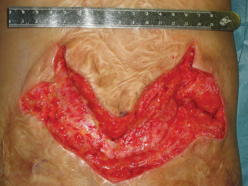
This is the first case report of long-term follow-up after applying the autologous cultured dermal substitute to establish the wound bed before split skin graft. The results suggest that application of autologous cultured cultured dermal substitute contributes to establish the high-quality wound bed for skin graft. Split-thickness skin grafts (STSGs) are the gold standard for the treatment of burn scar contracture. Young patients in particular may require additional skin grafts as they grow, and donor site for skin grafts may be limited. We applied autologous cultured dermal substitutes (CDSs) that are expected to establish a high-quality wound bed to allow thin STSGs. This is the first report of follow-up after application of autologous CDS combined with thin STSG. A male neonate suffered third-degree burns (20% of the total body surface area) on the back. After 2 years, scar contracture of the gluteal regions were released and autologous CDS were applied. Five days after the treatment, a super thin (4-6/1,000 per inch) skin grafting was performed. After 3 years, scar contracture of the back was released and autologous CDS was applied for 2 weeks. Then a split-thick graft was harvested from the same donor site. Ten years after the last operation, the width of the skin graft on his back has extended from 5-8 cm. The contour of the grafted skin is soft, smooth, and can be pinched. This long-term result shows the autologous CDS can be expected to establish the high-quality wound bed that allows thin STSG.
Figures


References
-
- McDonald WS, Deitch EA. Hypertrophic skin grafts in burned patients: a prospective analysis of variables. J Trauma. 1987;27:147. - PubMed
-
- Fujimori Y, Ueda K, Fumimoto H, et al. Skin regeneration for children with burn scar contracture using autologous cultured dermal substitutes and superthin auto-skin grafts: preliminary clinical study. Ann Plast Surg. 2006;57:408. - PubMed
-
- Kuroyanagi Y, Kubo K, Matsui H, et al. Establishment of banking system for allogeneic cultured dermal substitute. Artif Organs. 2004;28:13. - PubMed
-
- Hasegawa T, Suga Y, Mizoguchi M, et al. Clinical trial of allogeneic cultured dermal substitute for the treatment of intractable skin ulcers in 3 patients with recessive dystrophic epidermolysis bullosa. J Am Acad Dermatol. 2004;50:803. - PubMed
-
- Kubo K, Kuroyanagi Y. Development of a cultured dermal substitute composed of a spongy matrix of hyaluronic acid and atelo-collagen combined with fibroblasts: cryopreservation. Artif Organs. 2004;28:182. - PubMed
Publication types
LinkOut - more resources
Full Text Sources