

doi: 10.1016/j.vgie.2018.06.007.
eCollection 2018 Oct.
Endoscopic removal of a giant double-headed fibrovascular esophageal polyp
Affiliations
- PMID: 30276348
- PMCID: PMC6162200
- DOI: 10.1016/j.vgie.2018.06.007
Item in Clipboard
Endoscopic removal of a giant double-headed fibrovascular esophageal polyp
VideoGIE.
.
No abstract available
Figures
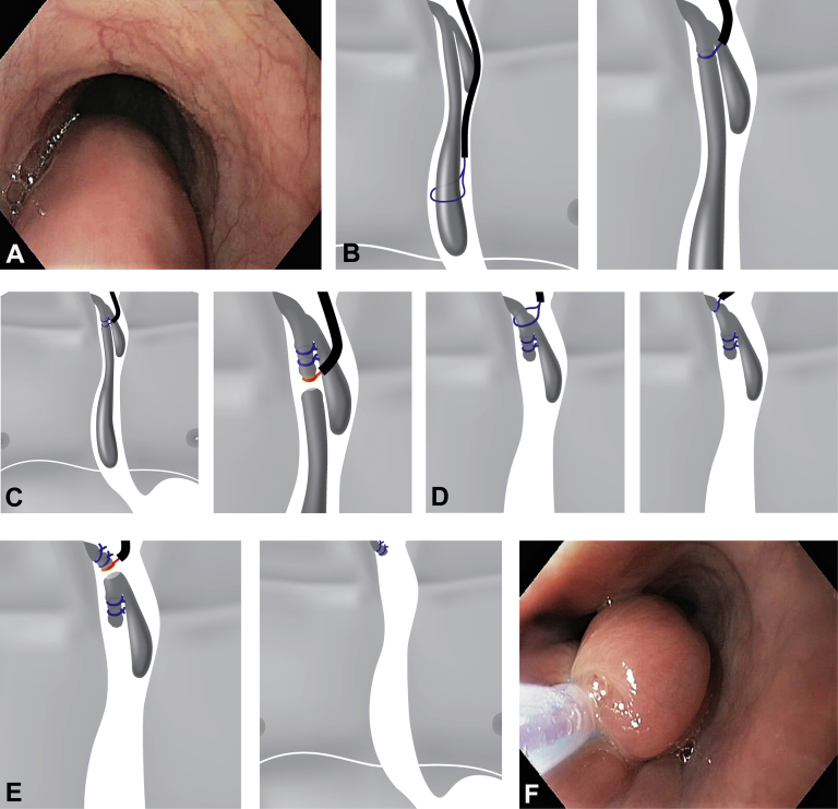
A, Upper-GI endoscopic view of giant fibrovascular polyp arising from the upper esophagus just near the level of the cricopharyngeus. Note huge submucosal appearance that occupies approximately the whole esophagus and has a smooth overlying mucosa of the body. B-E, General scheme of the removal of the giant esophageal fibrovascular polyp. B, C, Two endoloops, one after another, applied at the level of the ramification of the polyp body. The first portion of the polyp is cut. D, E, Another 2 endoloops are placed at the very base of the stalk and securely tightened. The remaining portion of the polyp is resected. F, First nylon endoloop is placed at the level of the ramification of the polyp body and tightened securely. The scope is then exchanged for a double-channel therapeutic gastroscope. Grasping forceps are used to manipulate the polyp in view of the difficulty of placing the edematous polyp through the second loop. G, Resection of the first 75-mm portion of the giant fibrovascular polyp above 2 endoloops in endocut mode with the 25-mm electrosurgical snare. H, The second loop is applied right over the first loop and tightened strongly, mainly to prevent possible profuse bleeding and perforation. The second portion of the polyp is removed above the loops. Resected giant fibrovascular polyp. I, Overall appearance of polyp. Total length, 13,5 cm; distal head, 32 mm in diameter with 2 ulcerations on the apex; proximal head, 25 mm in diameter. J, Cross-section at the level of the base of the polyp, 18 mm in diameter (note the large vessels). K, Ligated 10-mm stalk stump of the polyp left in place. L, M, Histologic appearance showing mixture of fibrous and adipose tissues accompanied by abundant network of large vessels, covered by normal squamous epithelium. (L, H&E, orig. mag. ×5; M, H&E, orig. mag. ×20.) N, Surveillance gastroscopic view at 18 months revealing no changes of esophageal wall and mucosa, with no evidence of recurrence.
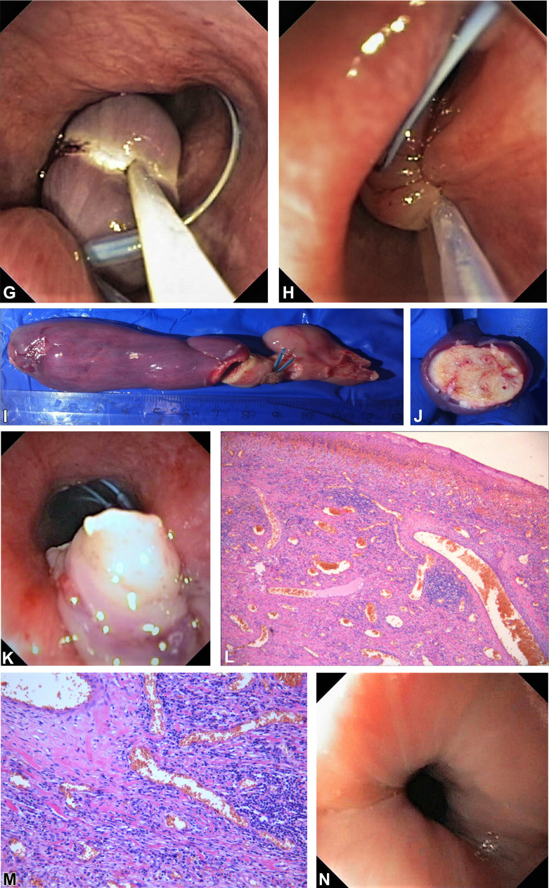
A, Upper-GI endoscopic view of giant fibrovascular polyp arising from the upper esophagus just near the level of the cricopharyngeus. Note huge submucosal appearance that occupies approximately the whole esophagus and has a smooth overlying mucosa of the body. B-E, General scheme of the removal of the giant esophageal fibrovascular polyp. B, C, Two endoloops, one after another, applied at the level of the ramification of the polyp body. The first portion of the polyp is cut. D, E, Another 2 endoloops are placed at the very base of the stalk and securely tightened. The remaining portion of the polyp is resected. F, First nylon endoloop is placed at the level of the ramification of the polyp body and tightened securely. The scope is then exchanged for a double-channel therapeutic gastroscope. Grasping forceps are used to manipulate the polyp in view of the difficulty of placing the edematous polyp through the second loop. G, Resection of the first 75-mm portion of the giant fibrovascular polyp above 2 endoloops in endocut mode with the 25-mm electrosurgical snare. H, The second loop is applied right over the first loop and tightened strongly, mainly to prevent possible profuse bleeding and perforation. The second portion of the polyp is removed above the loops. Resected giant fibrovascular polyp. I, Overall appearance of polyp. Total length, 13,5 cm; distal head, 32 mm in diameter with 2 ulcerations on the apex; proximal head, 25 mm in diameter. J, Cross-section at the level of the base of the polyp, 18 mm in diameter (note the large vessels). K, Ligated 10-mm stalk stump of the polyp left in place. L, M, Histologic appearance showing mixture of fibrous and adipose tissues accompanied by abundant network of large vessels, covered by normal squamous epithelium. (L, H&E, orig. mag. ×5; M, H&E, orig. mag. ×20.) N, Surveillance gastroscopic view at 18 months revealing no changes of esophageal wall and mucosa, with no evidence of recurrence.
LinkOut - more resources
Full Text Sources
