Observational Study
. 2019 Apr;29(4):1912-1921.
doi: 10.1007/s00330-018-5731-5. Epub 2018 Oct 1.

The value of post-mortem computed tomography of burned victims in a forensic setting

Affiliations
Observational Study

The value of post-mortem computed tomography of burned victims in a forensic setting

Henri M de Bakker et al. Eur Radiol. 2019 Apr.

Abstract

Objectives: Fire deaths are challenging fatalities for forensic pathologists, as the main question of whether death was due to the fire or not needs to be answered. In this retrospective study, we assessed whether post-mortem computed tomography (PMCT) has an added value prior to a forensic autopsy of burned victims.

Methods: From 2008 to 2016, a PMCT was performed in 50 burned corpses prior to a complete forensic autopsy. In retrospect, all 50 PMCT scans were systematically assessed by a forensically experienced radiologist, masked from the autopsy reports. Subsequently, the PMCT findings were compared with the autopsy reports.

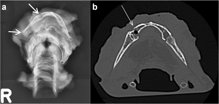
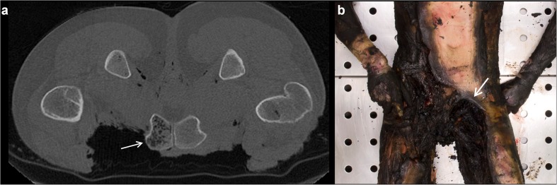
Results: Heat fractures, contractions and destruction of extremities, subcutaneous emphysema and post-mortem gas collections were easier to detect by PMCT compared to autopsy. Alterations by penetrating and blunt trauma and the presence of foreign bodies were easy to detect by PMCT as well by autopsy. PMCT was, however, not successful in detecting signs of vitality during the fire, detection of superficial thermal injuries and to answer the main question of the forensic autopsy, which is to investigate the cause of death.

Conclusions: PMCT prior to autopsy is a valuable add-on in the post-mortem forensic investigation of burned victims for detection of hidden signs of trauma, gas collections and foreign bodies. However, since PMCT cannot answer the two main questions in forensic examination-determining the cause of death and detecting signs of vitality during the fire-it cannot replace an autopsy.

Key points: • Post-mortem CT (PMCT) in burned victims shows hidden signs of trauma. • Foreign bodies and gas collections can easily be detected. • Cause of death and vitality signs cannot be assessed by PMCT.

Keywords: Burns; Forensic medicine; Forensic pathology; Radiology.


Conflict of interest statement

Guarantor

The scientific guarantor for this manuscript is Bernadette S. de Bakker, MD PhD.

Conflict of interest

The authors of this manuscript declare no relationships with any companies, whose products or services may be related to the subject matter of the article.

Statistics and biometry

No complex statistical methods were necessary for this paper.

Informed consent

Written informed consent was not required for this study because it concerned a retrospective study with anonymised data from deceased patients.

Ethical approval

Ethical Statement: The database contained anonymised patient data. Approval by a medical ethical committee for this anonymised retrospective investigation in deceased patients is not required to perform this type of study in The Netherlands.

Study subjects or cohorts overlap

Some study subjects or cohorts have been previously reported in de Bakker et al [6]. This methodological paper describes how we incorporated all forensic cases into a database for further research. None of the cases used in the current manuscript have been previously described, mentioned or presented.

Methodology

• Retrospective

• Observational

• Performed at one institution

Figures

Fig. 1
Fig. 1
Typical post-mortem CT findings in the head exposed to fire. a, b Axial images. In both cases, a dural tear and herniated brain tissue can be seen (single arrow). The case in (b) shows also an intracranial heat haematoma (double arrow). c Picture of an opened skull with heat haematoma (arrows) during autopsy. d, e Axial images. In both cases, heat fractures can be seen in the outer table (arrows) on locations where all soft tissue are burned away, resulting in separation of inner and outer table (split diploë sign). The case in (e) shows also a bilateral intracranial heat haematoma; *air-fluid level. f Enlarged section of (e). The arrows indicate the split diploë sign
Fig. 2
Fig. 2
Radiology of the explanted hyoid-larynx complex from a body exposed to fire. a X-ray shows fractures of the hyoid bone body and the greater horn on the right side (arrows). b Same case as in (a). Axial CT image of the body and greater horns of the hyoid bone. The fracture of the hyoid body is indicated by the white arrow. Histology showed haematomas at the fracture sites, which indicate that the person was alive when trauma to the neck was sustained
Fig. 3
Fig. 3
Examples of dense border sign on CT and at autopsy. a Axial image of a burned thorax. A ‘dense border sign’ can be seen at the surface of the lung that was exposed to fire (arrows). This is due to shrinkage and loss of fluid of the exposed tissue [3]. Note how the lungs are relatively intact in contrast to the absent burned surrounding structures. b Pathological specimen of the lung from the case presented in (a), showing a partly burned surface (arrows) that was in direct contact with the fire. c Axial image of a burned abdomen. A subtle dense border sign can be seen at part of the liver surface that was exposed to fire (arrows). See (d) for the pathological specimen of this case. d Pathological specimen of the liver from the case presented in (c), showing a partly burned surface (arrow) that was in direct contact with the fire, and a relatively normal part of the liver that was covered and protected from the fire by the body wall
Fig. 4
Fig. 4
Drug packages in a burned body. a Axial image of a body almost completely destructed by fire. Note the relative sparing of the drug packages (arrows). b Reconstructed sagittal image of the same case as in (a). Drug packages are indicated by arrows. c Image taken during autopsy of the carbonised body of the victim presented in (a) and (b). Note the light-coloured foreign body as small detail in the blackened tissue at autopsy. d Overview of the relatively unharmed drug packages that were recovered from the carbonised body presented in (ac) during autopsy
Fig. 5
Fig. 5
Typical proximal displacement of the patella in the left knee exposed to fire. a Anteroposterior 3D reconstruction of a CT of the lower extremities of a body exposed to fire, presenting patellar displacement. b Enlarged part of the 3D-CT scan in (a). Note the proximal displacement of the patella of the left knee (arrow). The patellar ligament is burned through or ruptured by the force of the shrinking rectus femoris muscle. c The proximally displaced and blackened left patella (black arrow) could be identified at autopsy. Its normal location is indicated by the dashed arrow. Typical flexing of the joints in legs and feet that were exposed to fire can be appreciated. Note also the coverage of the right patella by soft tissues, which prevents the patella from proximal displacement
Fig. 6
Fig. 6
Example of mottled lucencies in a burned body. a Axial image of the pubic bone, scanned in prone position as it was found at the crime scene. The left side of the pubic bone, not covered by soft tissues, contains mottled lucencies inside of the bone (arrow), whereas the right pubic bone appears normal [3]. b The same case as in (a) at autopsy. Ventral view of the pelvic region. Forearms and hands are also visible. The partially intact abdominal skin of the victim suggests a prone position during the fire. Note the heavily burned left groin (arrow) compared to the right groin that is still more or less covered by soft tissues

References

    1. Saukko PK, Knight B (2015) Knight's Forensic pathology, 4th edn, CRC Press, Boca Raton
    1. Kasahara S, Makino Y, Hayakawa M, Yajima D, Ito H, Iwase H. Diagnosable and non-diagnosable causes of death by postmortem computed tomography: a review of 339 forensic cases. Leg Med (Tokyo) 2012;14:239–245. doi: 10.1016/j.legalmed.2012.03.007. - DOI - PubMed
    1. Levy AD, Harcke HT, Getz JM, Mallak CT. Multidetector computed tomography findings in deaths with severe burns. Am J Forensic Med Pathol. 2009;30:137–141. doi: 10.1097/PAF.0b013e3181879cc9. - DOI - PubMed
    1. O'Donnell C, Iino M, Mansharan K, Leditscke J, Woodford N. Contribution of postmortem multidetector CT scanning to identification of the deceased in a mass disaster: Experience gained from the 2009 Victorian bushfires. Forensic Sci Int. 2011;205:15–28. doi: 10.1016/j.forsciint.2010.05.026. - DOI - PubMed
    1. Thali MJ, Yen K, Schweitzer W, et al. Virtopsy, a new imaging horizon in forensic pathology: virtual autopsy by postmortem multislice computed tomography (MSCT) and magnetic resonance imaging (MRI)—a feasibility study. J Forensic Sci. 2003;48:386–403. - PubMed