. 2019 Jan;111(1):131-144.
doi: 10.1111/mmi.14143. Epub 2018 Oct 21.

The small protein MgtS and small RNA MgrR modulate the PitA phosphate symporter to boost intracellular magnesium levels

Affiliations

The small protein MgtS and small RNA MgrR modulate the PitA phosphate symporter to boost intracellular magnesium levels

Xuefeng Yin et al. Mol Microbiol. 2019 Jan.

Abstract

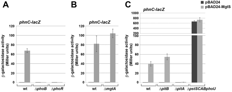
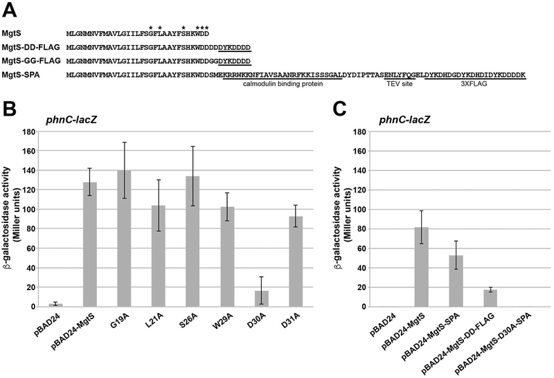
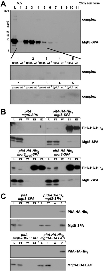
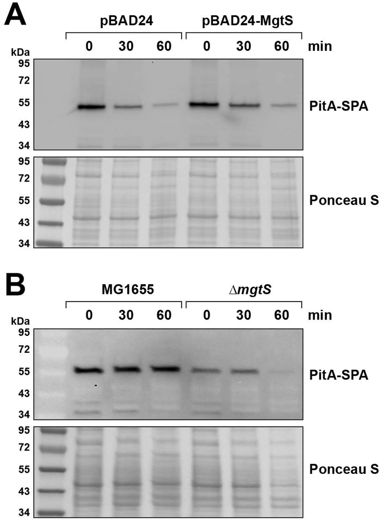
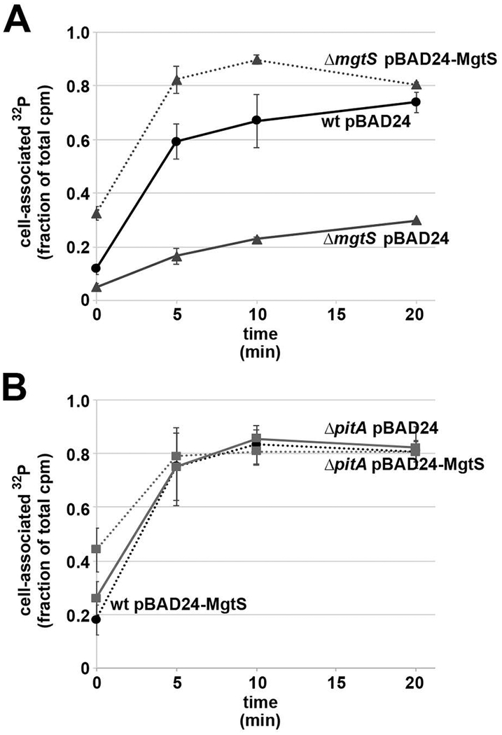
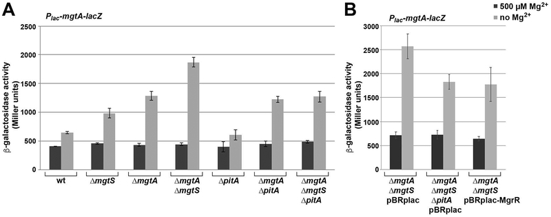
In response to low levels of magnesium (Mg2+ ), the PhoQP two component system induces the transcription of two convergent genes, one encoding a 31-amino acid protein denoted MgtS and the second encoding a small, regulatory RNA (sRNA) denoted MgrR. Previous studies showed that the MgtS protein interacts with and stabilizes the MgtA Mg2+ importer to increase intracellular Mg2+ levels, while the MgrR sRNA base pairs with the eptB mRNA thus affecting lipopolysaccharide modification. Surprisingly, we found overexpression of the MgtS protein also leads to induction of the PhoRB regulon. Studies to understand this activation showed that MgtS forms a complex with a second protein, PitA, a cation-phosphate symporter. Given that the additive effect of ∆mgtA and ∆mgtS mutations on intracellular Mg2+ concentrations seen previously is lost in the ∆pitA mutant, we suggest that MgtS binds to and prevents Mg2+ leakage through PitA under Mg2+ -limiting conditions. Consistent with a detrimental role of PitA in low Mg2+ , we also observe MgrR sRNA repression of PitA synthesis. Thus, PhoQP induces the expression of two convergent small genes in response to Mg2+ limitation whose products act to modulate PitA at different levels to increase intracellular Mg2+ .


Figures

Fig. 1.
Fig. 1.. MgtS induction of the PhoRB target gene phnC is pitA-dependent.
A. MgtS acts on the PhoRB two-component system to induce phnC expression. Strains with ΔmgtS (GSO797), ΔmgtS ΔphoB (GSO798) and ΔmgtS ΔphoR (GSO799) with a chromosomal PphnC-lacZ fusion and carrying the pBAD24 vector control or pBAD24-MgtS plasmid were inoculated into LB medium, grown to OD600 ~0.5 and treated with 0.2% arabinose for 30 min. Culture aliquots (1 mL) were collected for measurement of β-galactosidase activity. B. The induction of phnC expression by MgtS is not dependent on mgtA. Strains with ΔmgtS (GSO797) and ΔmgtS ΔmgtA (GSO800) with a chromosomal PphnC-lacZ fusion and carrying the pBAD24 vector control or pBAD24-MgtS plasmid were assayed for β-galactosidase activity as described above. C. The induction of phnC expression by MgtS requires pitA. Strains with ΔmgtS (GSO797), ΔmgtS ΔpitB (GSO802), ΔmgtS ΔpitA (GSO801), and ΔmgtS ΔpstSCAB-phoU (GSO803) with a chromosomal PphnC-lacZ fusion and carrying the pBAD24 vector control or pBAD24-MgtS plasmid were assayed for β-galactosidase activity as described above. For each strain, the enzyme activity reported is the average of three independent trials, and the error bars represent one SD.
Fig. 2.
Fig. 2.. D30A and MgtS-DD-FLAG derivatives of MgtS are defective for phnC-lacZ induction.
A. The sequence of MgtS and the different tagged derivatives. Asterisks indicate the mutated residues. B. A strain with ΔmgtS (GSO797) and a chromosomal PphnC-lacZ fusion carrying pBAD24, pBAD24-MgtS or indicated mutant derivatives were assayed for β-galactosidase activity as described for Fig. 1A. C. A strain with ΔmgtS (GSO797) and a chromosomal PphnC-lacZ fusion carrying pBAD24, pBAD24-MgtS or indicated tagged derivatives were assayed for β-galactosidase activity as described for Fig. 1A. Note the pBAD24 and pBAD-MgtS samples were the same as those in Fig 1B. For each strain, the enzyme activity reported is the average of three independent trials, and the error bars represent one SD.
Fig. 3.
Fig. 3.. MgtS-SPA associates with the PitA phosphate transporter.
A. Chemical crosslinking reveals a high molecular weight complex containing MgtS-SPA. The MgtS-D30A-SPA mutant does not form the complex. The complex also is not detected in the absence of PitA. Strains that were either pitA+ (MG1655) or ΔpitA (GSO815) and carried pBAD24-MgtS-SPA or pBAD24-MgtS-D30A-SPA plasmids were grown in LB medium to OD600 ~0.5 and treated with 0.2% arabinose for 7.5 min. Cells were then lysed and the whole cell lysate was separated by 5%−25% sucrose gradient sedimentation. Fractions recovered from gradient sedimentation were crosslinked with 25 mM DSS for 30 min prior to SDS-PAGE analysis and detection with α-FLAG antibody. B. Wild type MgtS-SPA but not MgtS-D30A-SPA mutant co-purifies with PitA-HA-His6. Strains carrying chromosomal alleles of mgtS-SPA (GSO767), mgtS-SPA pitA-HA-His6 (GSO804), MgtS-D30A-SPA (GSO805), or mgtS-D30A-SPA pitA-HA-His6 (GSO806) were grown in LB medium to OD600 ~0.5, collected, and lysed. The whole cell lysate (L) was applied onto a Ni-NTA column and flow-through (FT), wash (W) and two step-wise eluates (E1 and E2) were collected and analyzed by SDS-PAGE. MgtS-SPA and PitA-HA-His6 were detected by using α-FLAG antibody and α-HA antibody, respectively. C. MgtS-SPA but not MgtS-DD-FLAG co-purifies with PitA-HA-His6. Wild type E. coli (MG1655) carrying pBAD24 derivatives expressing PitA-HA-His6, MgtS-DD-FLAG or MgtS-SPA were grown in LB medium to OD600 ~0.5 and induced with 0.2% arabinose for 30 min, collected, and resuspended in lysis buffer. Wild type or PitA-HA-His6-expressing cells were mixed with those expressing MgtS-DD-FLAG or MgtS-SPA in a 2:3 ratio, homogenized, and applied to a Ni-NTA column for co-purification. After analysis by SDS-PAGE, MgtS-SPA and MgtS-DD-FLAG were detected using α-FLAG antibody and PitA-HA-His6 was detected using α-HA antibody.
Fig. 4.
Fig. 4.. MgtS slightly increases PitA-SPA levels.
A. Strains expressing a chromosomally-encoded SPA-tagged PitA (GSO807) and carrying pBAD24 or pBAD24-MgtS plasmids were grown in LB medium to OD600 ~0.1 whereupon an aliquot was taken (0 min). The remaining cells were induced with 0.2% arabinose and aliquots were taken at the indicated times. B. Strains expressing a chromosomally-encoded SPA-tagged PitA in a wild type (GSO807) or ΔmgtS (GSO820) background grown to OD600 ~0.1 in N-minimal medium with 500 μM Mg2+, was washed with and resuspended in N-minimal medium without added Mg2+, whereupon samples were taken at the indicated times. We found the ΔmgtS pitA-SPA strain grew poorly in N-minimal medium without added Mg2+, which likely accounts for the reduced Ponceau staining at the 60 min time point. For all time points, aliquots were pelleted, resuspended to OD600 ~10 in SDS loading buffer, and separated by SDS-PAGE for immunoblot analysis using anti–FLAG antibodies.
Fig. 5.
Fig. 5.. MgtS increases cell-associated 32P levels
A. Wild type (GSO770) carrying pBAD24 and ΔmgtS (GSO772) carrying pBAD24 or pBAD24- MgtS plasmids were transferred to in N minimal media without added Mg2+, induced for 30 min, washed and resuspended in Tris-buffered saline. B. Wild type (GSO770) carrying pBAD24 and ΔpitA (GSO816) carrying pBAD24 or pBAD24- MgtS plasmids were transferred to in N minimal media without added Mg2+, induced for 30 min, washed and resuspended in Tris-buffered saline. For both sets of strains, 32P-potassium phosphate was added, and an aliquot removed (0 min). The samples were incubated at 37°C, and at the indicated time points, aliquots were removed for the determination of CPM/mL.
Fig. 6.
Fig. 6.. ΔpitA and MgrR sRNA overexpression eliminate additional induction of Mg2+-dependent fusion in ΔmgtA ΔmgtS background.
A. β-galactosidase activity was assayed for cultures of wild type (GSO770), ΔmgtS (GSO772), ΔmgtA (GSO808), ΔmgtA ΔmgtS (GSO809), ΔpitA (GSO810), ΔmgtA ΔpitA (GSO811), and ΔmgtA ΔmgtS ΔpitA (GSO812) carrying the chromosomal Plac-leadermgtA-lacZ fusion grown in N medium with 500 μM Mg2+ or without added Mg2+ and assayed for β-galactosidase as described (Wang et al., 2017). B. ΔmgtA ΔmgtS (GSO809) and ΔmgtA ΔmgtS ΔpitA (GSO812) carrying the chromosomal Plac-leadermgtA-lacZ fusion and containing pBR-plac or pBR-plac-MgrR as indicated were assayed for β-galactosidase activity as described for Fig. 5A. For each strain, the enzyme activity reported is the average of three independent trials, and the error bars represent one SD.
Fig. 7.
Fig. 7.. pitA is repressed by the MgrR sRNA.
A. Chimeras (indicated in red and dark blue) found in RIL-Seq experiments (Melamed et al., 2016) encompass region (light blue) of base pairing predicted between pitA mRNA and MgrR using IntaRNA (Mann et al., 2017) in December 2017. Numbering is with respect to start codon of the pitA ORF and +1 of MgrR. B. MgrR overexpression leads to reduced PitA-SPA levels. Strains containing a chromosomally-encoded PitA-SPA (GSO807) and pBRplac or pBRplac-MgrR plasmids were grown in LB medium with 1 mM IPTG to OD600 ~0.5 and ~1.0, whereupon aliquots were taken. A strain expressing untagged PitA (MG1655) and GSO807 without a plasmid grown to OD600 ~0.5 were used as negative and positive controls, respectively. For all samples, aliquots were pelleted, resuspended to OD600 ~10 in SDS loading buffer, and separated by SDS-PAGE for immunoblot analysis using anti–FLAG antibodies. C. MgrR overexpression leads to reduced pitA mRNA levels. Wild type MG1655 carrying pBRplac or pBRplac-MgrR plasmids and ΔpitA (GSO816) carrying pBRplac were grown in LB medium with 1 mM IPTG to OD600 ~0.5 whereupon total RNA was isolated and separated on an agarose gel for northern blot detection with probes against the pitA mRNA, MgrR sRNA, and 5S rRNA.

References

    1. Adams PP, Flores Avile C, Popitsch N, Bilusic I, Schroeder R, Lybecker M, and Jewett MW (2017) In vivo expression technology and 5’ end mapping of the Borrelia burgdorferi transcriptome identify novel RNAs expressed during mammalian infection. Nucleic Acids Res. 45: 775–792. - PMC - PubMed
    1. Anderson DM, Makarewich CA, Anderson KM, Shelton JM, Bezprozvannaya S, Bassel-Duby R, and Olson EN (2016) Widespread control of calcium signaling by a family of SERCA-inhibiting micropeptides. Sci. Signal 9: ra119. - PMC - PubMed
    1. Azam MS, and Vanderpool CK (2018) Translational regulation by bacterial small RNAs via an unusual Hfq-dependent mechanism. Nucleic Acids Res. 46: 2585–2599. - PMC - PubMed
    1. Bader MW, Sanowar S, Daley ME, Schneider AR, Cho U, Xu W, Klevit RE, Le Moual H, and Miller SI (2005) Recognition of antimicrobial peptides by a bacterial sensor kinase. Cell 122: 461–472. - PubMed
    1. Baym M, Lieberman TD, Kelsic ED, Chait R, Gross R, Yelin I, and Kishony R (2016) Spatiotemporal microbial evolution on antibiotic landscapes. Science 353: 1147–1151. - PMC - PubMed

Publication types

MeSH terms