. 2019 Jan;81(1):439-453.
doi: 10.1002/mrm.27420. Epub 2018 Sep 18.

Scan-specific robust artificial-neural-networks for k-space interpolation (RAKI) reconstruction: Database-free deep learning for fast imaging

Affiliations

Scan-specific robust artificial-neural-networks for k-space interpolation (RAKI) reconstruction: Database-free deep learning for fast imaging

Mehmet Akçakaya et al. Magn Reson Med. 2019 Jan.

Abstract

Purpose: To develop an improved k-space reconstruction method using scan-specific deep learning that is trained on autocalibration signal (ACS) data.

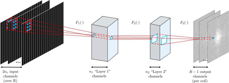
Theory: Robust artificial-neural-networks for k-space interpolation (RAKI) reconstruction trains convolutional neural networks on ACS data. This enables nonlinear estimation of missing k-space lines from acquired k-space data with improved noise resilience, as opposed to conventional linear k-space interpolation-based methods, such as GRAPPA, which are based on linear convolutional kernels.

Methods: The training algorithm is implemented using a mean square error loss function over the target points in the ACS region, using a gradient descent algorithm. The neural network contains 3 layers of convolutional operators, with 2 of these including nonlinear activation functions. The noise performance and reconstruction quality of the RAKI method was compared with GRAPPA in phantom, as well as in neurological and cardiac in vivo data sets.

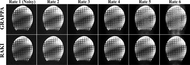
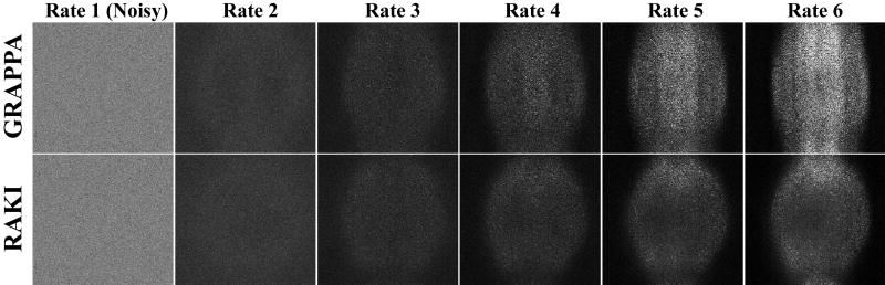
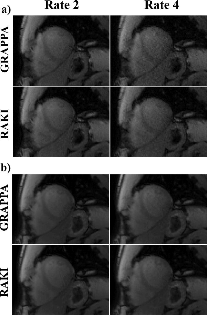
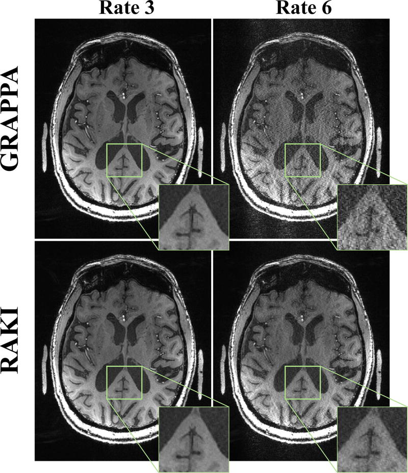
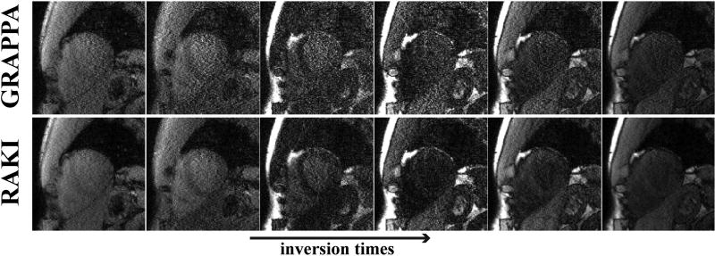
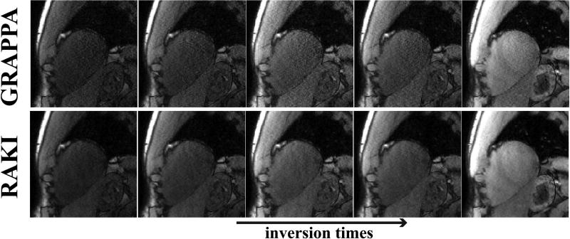
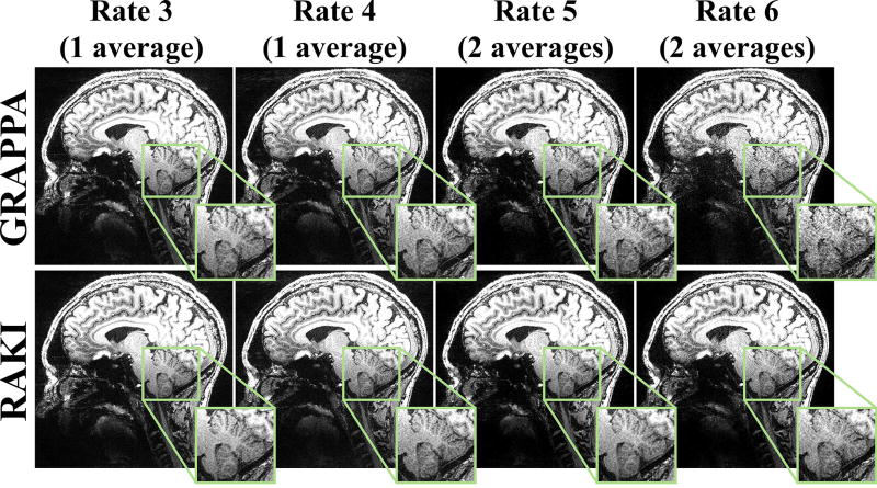
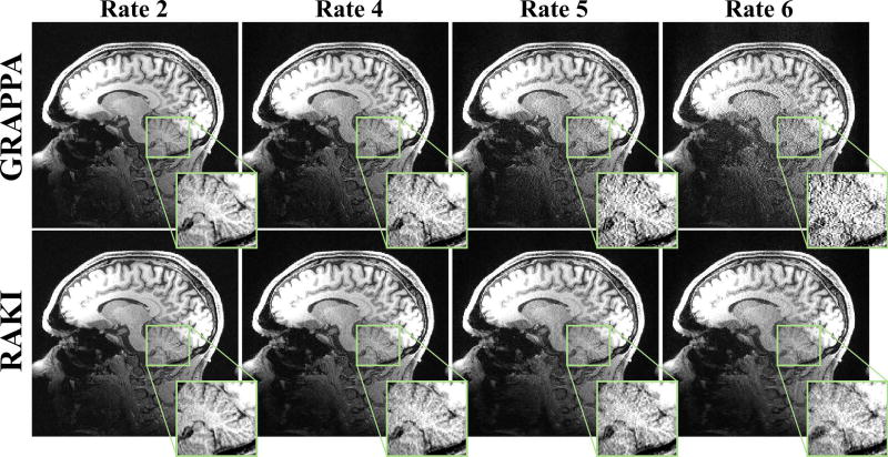
Results: Phantom imaging shows that the proposed RAKI method outperforms GRAPPA at high (≥4) acceleration rates, both visually and quantitatively. Quantitative cardiac imaging shows improved noise resilience at high acceleration rates (rate 4:23% and rate 5:48%) over GRAPPA. The same trend of improved noise resilience is also observed in high-resolution brain imaging at high acceleration rates.

Conclusion: The RAKI method offers a training database-free deep learning approach for MRI reconstruction, with the potential to improve many existing reconstruction approaches, and is compatible with conventional data acquisition protocols.

Keywords: accelerated imaging; convolutional neural networks; deep learning; image reconstruction; k-space interpolation; nonlinear estimation; parallel imaging.


Figures

Figure 1
Figure 1
The three layer network structure used in this study for the convolutional neural network. The first layer, F1 (·), takes in the sub-sampled zero-filled k-space of size nx×ny×2nc, as embedded into the real field. The convolutional filters in this layer, w1 are of size b1x×b1y×2nc×n1. This is followed by a rectified linear unit (ReLU) operation. The second layer of our network, F2 (·), takes in the output of the first layer and applies convolutional filters, denoted by w2, are of size b2x×b2y×n1×n2. This layer also includes a ReLU operation. These two layers non-linearly combine the acquired k-space lines. The final layer of the network, F3 (·), produces the desired reconstruction output by applying convolutional filters, w3, of size b3x×b3y×n2×nout. In our implementation all missing lines per coil (over reals) is estimated simultaneously, therefore nout = R − 1.
Figure 2
Figure 2
Results of phantom experiments. Images reconstructed using GRAPPA (top row) and the proposed RAKI (bottom row) reconstructions from noisy data sets for various acceleration rates, as well as a fully-sampled noisy reference image. Starting with rate 4, the noise performance of RAKI improves upon that of GRAPPA. RAKI reconstructions at acceleration rates 4 and 5 show visually improved noise properties over GRAPPA, which has high levels of noise amplification.
Figure 3
Figure 3
The difference images for the reconstructions in Figure 2, as compared to the original acquisition without additional noise, confirm the observations regarding the improved noise resilience of RAKI. NMSE quantification is also consistent with these observations: 0.65, 0.51, 0.51, 0.66 and 0.89 for GRAPPA versus 0.65, 0.51, 0.46, 0.47 and 0.52 for RAKI from rates 2 to 6.
Figure 4
Figure 4
Representative T1-weighted images from SAPPHIRE T1 mapping acquired at the conventional resolution of 1.7×1.7 mm2. (a) Images with low acquisition SNR, corresponding to the shortest inversion time, at the acquisition acceleration rate of 2, and a retrospective acceleration rate of 4. At rate 4, the noise performance improvement of RAKI over GRAPPA becomes apparent. (b) Images with high acquisition SNR, where the differences between RAKI and GRAPPA are visually minor even at the acceleration rate of 4, with a slight improvement in the left ventricle blood pool for RAKI. A quantitative analysis of the resulting T1 maps indicate that comparable quantification is observed for the two reconstructions, except for a 10% reduced noise variability with RAKI at rate 4.
Figure 5
Figure 5
6 T1-weighted images, corresponding to the shortest inversion times, acquired using the SAPPHIRE sequence at a high spatial resolution of 1.1×1.1 mm2, and acceleration rate 5. At this high acceleration rate, the noise improvement of RAKI over GRAPPA is observable for all images. A quantitative analysis of the resulting T1 maps indicate that the spatial variability of the myocardial T1 is improved by 37% using RAKI versus GRAPPA.
Figure 6
Figure 6
5 T1-weighted images, corresponding to the longest inversion times, from the same dataset depicted in Figure 5. At this high acceleration rate, the noise improvement of RAKI is observable for all images. Quantitative analysis of T1 map spatial variability shows 37% improvement using RAKI versus GRAPPA.
Figure 7
Figure 7
A central slice of the high-resolution (0.6 mm isotropic) 7T MPRAGE acquisition, both at the acquisition acceleration rate of 3, as well as at a retrospective acceleration rate of 6. At rate 3, GRAPPA (top) and RAKI (bottom) methods both successfully reconstruct the image with little residual artifacts. At rate 6, GRAPPA reconstruction suffers heavily from noise amplification, whereas the proposed RAKI reconstruction exhibits improved noise tolerance without blurring artifacts.
Figure 8
Figure 8
A central slice of the high-resolution (0.6 mm isotropic) MPRAGE acquisition at 7T, acquired at R = 3, 4, 5 and 6, where the first two rates were acquired with one average, and the latter two were acquired with two averages. Thus, the only SNR penalty between R = 3 and 6 is due to differences in coil encoding. The results show that at this high SNR, GRAPPA (top) and RAKI (bottom) reconstruct the images with little residual artifacts up to R = 4. At R = 5, slight differences between GRAPPA and RAKI can be observed, with the latter showing better noise performance. At R = 6, the difference is further pronounced. At the higher rates, RAKI has improved noise tolerance while exhibiting no blurring artifacts.
Figure 9
Figure 9
A central slice of the high-resolution (0.7 mm isotropic) MPRAGE acquisition at 3T, acquired at R = 2 and 5, as well as retrospective R = 4 and 6. At rates 2 and 4, GRAPPA (top) and RAKI (bottom) methods both successfully reconstruct the image with little residual artifacts. At rates 5 and 6, noise amplification becomes visible for GRAPPA reconstruction. At these rates, RAKI has improved noise tolerance and exhibits no blurring artifacts.

References

    1. Sodickson DK, Manning WJ. Simultaneous acquisition of spatial harmonics (SMASH): fast imaging with radiofrequency coil arrays. Magn Reson Med. 1997;38(4):591–603. - PubMed
    1. Pruessmann KP, Weiger M, Scheidegger MB, Boesiger P. SENSE: sensitivity encoding for fast MRI. Magn Reson Med. 1999;42(5):952–962. - PubMed
    1. Griswold MA, Jakob PM, Heidemann RM, Nittka M, Jellus V, Wang J, Kiefer B, Haase A. Generalized autocalibrating partially parallel acquisitions (GRAPPA) Magn Reson Med. 2002;47(6):1202–1210. - PubMed
    1. Lustig M, Pauly JM. SPIRiT: Iterative self-consistent parallel imaging reconstruction from arbitrary k-space. Magn Reson Med. 2010;64(2):457–471. - PMC - PubMed
    1. Meyer CH, Hu BS, Nishimura DG, Macovski A. Fast spiral coronary artery imaging. Magn Reson Med. 1992;28(2):202–213. - PubMed

Publication types

LinkOut - more resources