. 2019 Jan;9(1):82-95.
doi: 10.1158/2159-8290.CD-18-0168. Epub 2018 Oct 2.

Age-Related Changes in HAPLN1 Increase Lymphatic Permeability and Affect Routes of Melanoma Metastasis

Affiliations

Age-Related Changes in HAPLN1 Increase Lymphatic Permeability and Affect Routes of Melanoma Metastasis

Brett L Ecker et al. Cancer Discov. 2019 Jan.

Abstract

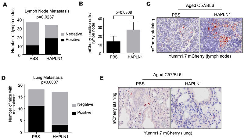
Older patients with melanoma have lower rates of sentinel lymph node (LN) metastases yet paradoxically have inferior survival. Patient age correlated with an inability to retain Technetium radiotracer during sentinel LN biopsy in more than 1,000 patients, and high Technetium counts correlated to better survival. We hypothesized that loss of integrity in the lymphatic vasculature due to extracellular matrix (ECM) degradation might play a role. We have implicated HAPLN1 in age-dependent ECM degradation in the dermis. Here, we queried whether HAPLN1 could be altered in the lymphatic ECM. Lymphatic HAPLN1 expression was prognostic of long-term patient survival. Adding recombinant HAPLN1 to aged fibroblast ECMs in vitro reduced endothelial permeability via modulation of VE-cadherin junctions, whereas endothelial permeability was increased following HAPLN1 knockdown in young fibroblasts. In vivo, reconstitution of HAPLN1 in aged mice increased the number of LN metastases, but reduced visceral metastases. These data suggest that age-related changes in ECM can contribute to impaired lymphatics. SIGNIFICANCE: Our studies reveal that changes in the stroma during aging may influence the way tumor cells traffic through the lymphatic vasculature. Aging may dictate the route of metastatic dissemination of tumor cells, and understanding these changes may help to reveal targetable moieties in the aging tumor microenvironment.See related commentary by Marie and Merlino, p. 19.This article is highlighted in the In This Issue feature, p. 1.


Conflict of interest statement

Conflict of Interest: The authors declare no potential conflicts of interest

Figures

Figure 1.
Figure 1.. Retention of technicium dye and patterns of metastatic dissemination correlate with age.
A. Consecutive melanoma patients (n=1081) were injected with 99mTc sulfur colloid in the region of the primary tumor and a gamma probe was used to quantify the signal intensity in the surgically resected sentinel lymph node (Spearman’s ρ=−0.30, p<0.001) B. Representative lymphoscintigraphy of Tc-99m sulfur colloid signal in young and aged melanoma patients; C. Survival curve adjusted for #of positive SLN, T-stage and ulceration, stratified by hottest counts above or below the median (HR 0.831, p=0.0014 95% CI 0.719-0.92); D. Kaplan-Meier analysis of distant metastatic-free survival in sentinel lymph node biopsy negative patients, stratified by patient age at diagnosis (n=1,649; log rank p<0.001); E. mCherry-labeled Yumm1.7 cells were injected into young or aged C57BL/6 mice and metastatic cells were identified in the draining inguinal lymph node by immunohistochemistry (bar=100μm). Number of cells were counted per lymph node and graphed (two-tailed unpaired t-test, p=0.0030); F. The mean number of metastases per high power field was similarly determined for lungs from the matching young and aged C57BL/6 mouse cohort (two-tailed unpaired t-test, p=0.0029).
Figure 2.
Figure 2.. Changes in lymphatic fibroblast ECM deposition according to age.
A. GSEA analysis for ECM fibril organization of lymph nodes from human melanoma patients (n=221; nominal p=0.008); B. In vitro extracellular matrix was produced by lymphatic fibroblasts isolated from young and aged donors and analyzed for the levels of fiber orientation by fibronectin immunofluorescence. The fiber distribution was determined by calculating the percent of fibers arranged in parallel for each acquired region (± 90° of the mode angle); each point in the corresponding dotplot represent the mean number of fibers orientated at each angle (paired bars represent the standard error of the mean); C. Analysis of extracellular matrix orientation produced in vitro by dermal fibroblasts from young or aged healthy donors; D. Schematic of experimental setup: Matrices derived from young and aged fibroblasts were reconstituted with HUVECs followed by incubation with Texas red dye in the upper transwell chamber. Permeability was determined by quantification of the fluorescence of the lower chamber after 30 minutes. E, Permeability of endothelial cells plated on young and aged matrices as measured by Texas red (two-tailed unpaired t-test: young vs. aged, p<0.001); F. Transwell permeability assay of HUVECs plated on acellular extracellular matrices produced by extracted aged fibroblasts treated with increasing doses of rHAPLN1 (ANOVA p=0.0032; two-tailed unpaired t-test: 0 ng vs. 5 ng, p=0.0205; 0 ng vs. 25 ng, p=0.0080); G. Transwell permeability assay of HUVECs plated on acellular extracellular matrices produced by extracted young fibroblasts with shHAPLN1 knockdown (ANOVA p=0.0004; two-tailed unpaired t-test: shempty vs. sh0501, p=0.0004; shempty vs. sh3400, p=0.0056); H. Quantification of GFP-labeled melanoma cell migration (48 hours) on cell derived matrices under different conditions (ANOVA p=0.0004; all pairwise comparisons by two-tailed t-tests, p<0.001).
Figure 3.
Figure 3.. Changes in cell-adhesion and integrin expression with age.
A. Representative VE-cadherin (red) confocal immunofluorescence of HUVECs plated on acellular matrices following extraction of young shEmpty and shHAPLN1 fibroblasts and aged fibroblasts treated with rHAPLN1 (25 ng/mL) or PBS (bar=100μm); B. Corresponding quantification of the signal intensity of VE-cadherin positive cellular adhesions between HUVECs; C. Relative expression by qtPCR of integrins (ITGB1, ITGB5, IGTA1, ITGA5) and CD44 of HUVECs on acellular matrices produced by young or aged fibroblasts (** p<0.01); D. Relative expression by qtPCR of integrins (ITGB1, ITGB5, IGTA1, ITGA5) and CD44 of HUVECs on acellular matrices produced by young fibroblasts following HAPLN1 knockdown (* p<0.05; ** p<0.01); E. Representative podoplanin (brown) and VE-cadherin (red) two-color immunohistochemistry of human sentinel lymph node specimens from primary cutaneous melanoma patients (n=16; bar=100μm); F. VE-cadherin signal quantification from human sentinel lymph nodes (n=16; two-tailed unpaired t-test, p=0.0003).
Figure 4.
Figure 4.. Effects of HAPLN1 on lymph node integrity.
A. Representative two-photon microscopy of pericapsular collagen structure of inguinal lymph node in young or aged C57BL/6 mice, and corresponding quantification; B, HAPLN1 mRNA expression as measured by RT-PCR. C. Representative two-photon microscopy of pericapsular collagen structure of inguinal lymph node in aged C57BL/6 mice treated with rHAPLN1 (100 ng twice weekly) or PBS; D. Quantification of young and aged lymphatic pericapsular extracellular matrix fiber orientation by collagen fluorescence. Each point in the corresponding dotplot represent the mean number of fibers orientated at each angle (paired bars represent the standard error of the mean). The fiber distribution was determined by calculating the percent of fibers arranged in parallel for each acquired region (±90° of the mode angle); the percent of fibers within 15° of the mode was compared between study arms (two-tailed unpaired t-test, p=0.0009); E. Quantification of lymphatic pericapsular extracellular matrix fiber orientation by collagen fluorescence after HAPLN1 treatment; F. Representative HAPLN1 immunohistochemistry of sentinel lymph node specimens of clinically node-negative melanoma patients (bar=100μm). Each sample was assigned a H-score that included the relative signal intensity and area of staining (n=30; two-tailed unpaired t-test, p=0.0310); G. Age-stratified TCGA analysis of HAPLN1 mRNA expression in regional lymphatic tissue of primary melanoma patients (n=192; two-tailed unpaired t-test, p=0.0324); H. Geiger counts of melanoma patients following sentinel lymph node biopsy with Tc-99m sulfur colloid injection, stratified by HAPLN1-positivity by immunohistochemistry staining (n=86; two-tailed unpaired t-test, p=0.0046); I. Kaplan-Meier survival function of non-metastatic human melanoma patients in the TCGA database, stratified by quartiles of regional lymph node HAPLN1 mRNA (n=192; log rank p<0.001).
Figure 5.
Figure 5.. In vivo effects of HAPLN1 on routes of metastatic dissemination.
A. mCherry-labeled yumm 1.7 cells were injected into aged C57BL/6 mice (n=18/arm) and the draining lymph nodes were treated with HAPLN1 (100 ng) or PBS. Tumor metastasis in the draining lymphatics were identified by immunohistochemical staining (chi squared p=0.0237); B. Lymphatic tumor burden was quantified (two-tailed unpaired t-test, p=0.0308); C. Representative immunohistochemistry of mCherry-positive metastasis (red) in draining lymphatics; D. Tumor metastasis in the lung in the identical mouse cohort were identified by immunohistochemical staining (chi squared p=0.0087); E. Representative immunohistochemistry of mCherry-positive metastasis (red) in the lungs.
Figure 6.
Figure 6.. Schematic representation of age-dependent changes in melanoma tumor progression.
Age-related changes in the peri-lymphatic stroma impair the integrity of lymphatic vessels and nodes and increase lymphatic permeability. Such differences may underlie the clinical observations of increased rates of in-transit disease and false negative sentinel lymph node biopsies.

Comment in

References

    1. Balch CM, Soong SJ, Gershenwald JE, Thompson JF, Reintgen DS, Cascinelli N, et al. Prognostic factors analysis of 17,600 melanoma patients: validation of the American Joint Committee on Cancer melanoma staging system. J Clin Oncol. 2001;19:3622–34. - PubMed
    1. Amin MB, Greene FL, Edge SB, Compton CC, Gershenwald JE, Brookland RK, et al. The Eighth Edition AJCC Cancer Staging Manual: Continuing to build a bridge from a population-based to a more “personalized” approach to cancer staging. CA Cancer J Clin. 2017;67:93–9. - PubMed
    1. Balch CM, Soong SJ, Gershenwald JE, Thompson JF, Coit DG, Atkins MB, et al. Age as a prognostic factor in patients with localized melanoma and regional metastases. Ann Surg Oncol. 2013;20:3961–8. - PMC - PubMed
    1. Lasithiotakis K, Leiter U, Meier F, Eigentler T, Metzler G, Moehrle M, et al. Age and gender are significant independent predictors of survival in primary cutaneous melanoma. Cancer. 2008;112:1795–804. - PubMed
    1. Morton DL. Overview and update of the phase III Multicenter Selective Lymphadenectomy Trials (MSLT-I and MSLT-II) in melanoma. Clin Exp Metastasis. 2012;29:699–706. - PMC - PubMed

Publication types