. 2019 Jan;9(1):64-81.
doi: 10.1158/2159-8290.CD-18-0193. Epub 2018 Oct 2.

Remodeling of the Collagen Matrix in Aging Skin Promotes Melanoma Metastasis and Affects Immune Cell Motility

Affiliations

Remodeling of the Collagen Matrix in Aging Skin Promotes Melanoma Metastasis and Affects Immune Cell Motility

Amanpreet Kaur et al. Cancer Discov. 2019 Jan.

Abstract

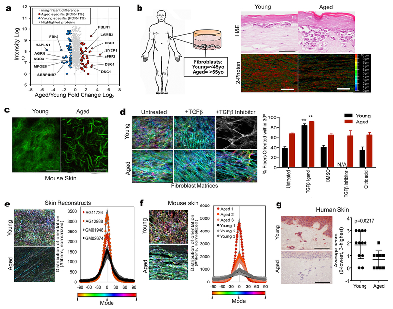
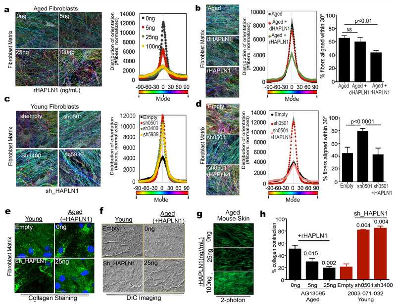
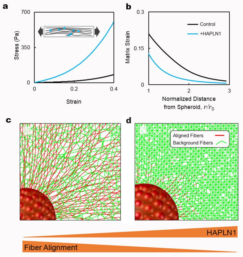
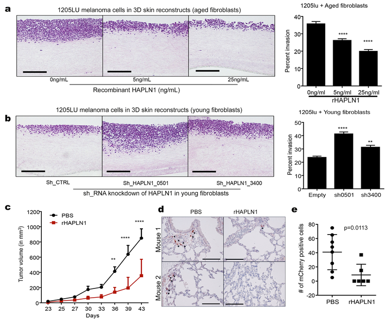
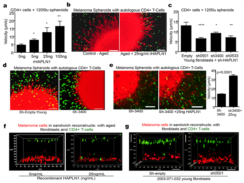
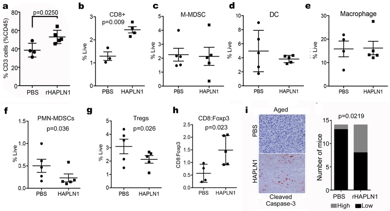
Physical changes in skin are among the most visible signs of aging. We found that young dermal fibroblasts secrete high levels of extracellular matrix (ECM) constituents, including proteoglycans, glycoproteins, and cartilage-linking proteins. The most abundantly secreted was HAPLN1, a hyaluronic and proteoglycan link protein. HAPLN1 was lost in aged fibroblasts, resulting in a more aligned ECM that promoted metastasis of melanoma cells. Reconstituting HAPLN1 inhibited metastasis in an aged microenvironment, in 3-D skin reconstruction models, and in vivo. Intriguingly, aged fibroblast-derived matrices had the opposite effect on the migration of T cells, inhibiting their motility. HAPLN1 treatment of aged fibroblasts restored motility of mononuclear immune cells, while impeding that of polymorphonuclear immune cells, which in turn affected regulatory T-cell recruitment. These data suggest that although age-related physical changes in the ECM can promote tumor cell motility, they may adversely affect the motility of some immune cells, resulting in an overall change in the immune microenvironment. Understanding the physical changes in aging skin may provide avenues for more effective therapy for older patients with melanoma. SIGNIFICANCE: These data shed light on the mechanochemical interactions that occur between aged skin, tumor, and immune cell populations, which may affect tumor metastasis and immune cell infiltration, with implications for the efficacy of current therapies for melanoma.See related commentary by Marie and Merlino, p. 19.This article is highlighted in the In This Issue feature, p. 1.


Conflict of interest statement

Conflict of Interest: The authors declare no potential conflicts of interest

Figures

Figure 1:
Figure 1:. The extracellular matrix is significantly altered during aging.
a) Secretome analysis from conditioned media from young and aged fibroblasts showing top overexpressed proteins in both and young and aged microenvironment. b) Dermal fibroblasts from healthy human donors were used to prepare skin reconstructs. Invasion and collagen deposition was assessed by H&E staining (upper panels; bar=100microns) as well as two-photon microscopy (bottom panels; bar=25microns). The color scale is indicative of the thickness of the collagen. c) C57/BL6 mouse skin was assessed for collagen composition using two-photon microscopy (bar=25 microns). d) Young and aged dermal fibroblasts were allowed to form matrices and color-coded for fiber alignment. More colors represent less alignment. Panel shown in black and white indicates no significant matrix. Results are also quantified along with controls. N/A indicates no significant matrix formed. e) Skin reconstructs were prepared using multiple young (GM01948, GM02674) and aged (AG11726, AG12988) fibroblasts and matrix alignment was measured. f) Young and aged C57/BL6 mice were injected with Yumm1.7 tumors and analyzed for matrix orientation at the skin/tumor interface. g) Normal human non-melanoma skin from young and aged donors was stained for expression of HAPLN1 and scored based on intensity (3-highest, 0-lowest/absent).
Figure 2:
Figure 2:. HAPLN1 loss in aging microenvironment promotes extracellular matrix remodeling.
a) Matrix production by aged fibroblasts was carried out in presence of various concentrations of recombinant HAPLN1, and alignment of the matrix was measured. b) Aged fibroblasts were treated with active or denatured HAPLN1 (25ng/ml), allowed to form matrices and matrix alignment was measured. c) Young fibroblasts with HAPLN1 knockdown were allowed to produce matrix and assessed for matrix production. d) Young fibroblasts with HAPLN1 knockdown were allowed to form matrices with or without presence of rHAPLN1 (25ng/ml) and assessed for matrix alignment. e) Matrices were produced by young fibroblasts with HAPLN1 knockdown and aged fibroblasts with rHAPLN1 treatment (25ng/ml). Matrices were assessed for Collagen I by immunofluorescence (bar=25microns). f) Skin reconstructs were prepared with young fibroblasts with HAPLN1 knockdown and aged fibroblasts with rHAPLN1 treatment (25ng/ml). Skin reconstructs were embedded in paraffin and imaged for collagen bundles using Differential Interference Contrast (DIC) microscopy. g) Aged C57/BL6 mice were treated intradermally with 100ng (50ng/mL) rHAPLN1 for 7 days, followed by two photon imaging for dermal collagen (bar=25microns). h) Collagen was embedded with aged fibroblasts treated with varying concentrations of rHAPLN1 and young fibroblasts with HAPLN1 knockdown and layered in 48 well plates and assessed for contractility over 3 days (ANOVA (young+ shHAPLN1) 0.0022, (aged+rHAPLN1) 0.0031). For all experiments, ANOVA post-hoc tests were performed using Bonferroni correction and the significance values are shown in the figures.
Figure 3.
Figure 3.. Chemo-Mechanical model for HAPLN1 restriction of tumor invasion into the extracellular matrix.
a) Stress-strain curves of fibrous networks with and without HAPLN1 obtained using stretch tests. The addition of HAPLN1 is modeled by the formation of crosslinks between nearby fibers. The inset shows a schematic of the fibrous networks in the stretch tests. The fibers and crosslinks are shown in black and red, respectively. b) The results from panel (a) are then used to inform our chemo-mechanical model about the mechanical behavior of fibrous matrices with and without HAPLN1. Strain in the matrix is plotted as a function of distance from the tumor spheroid. c) Fibers are aligned by the contractility of a tumor spheroid in the control case. The aligned and background fibers are displayed in red and green, respectively. Fibers are colored based on the stretching force present in the fibers. d) Reduced matrix deformation and fiber alignment are observed in the case where more fibers are crosslinked by HAPLN1. At the same level of strain, fewer fibers buckle in the matrix with HAPLN1 because crosslinks constrain the lateral bending of individual fibers, preventing the alignment of fibers due to cell contractility.
Figure 4:
Figure 4:. HAPLN1 decreases invasion and metastasis of melanoma cells.
a) Skin reconstructs were prepared with aged fibroblasts treated with varying concentrations of rHAPLN1 and invasion was calculated as a percent of reconstruct thickness (ANOVA p<0.0001, bar=100microns). b) Skin reconstructs prepared with young fibroblasts with HAPLN1 knockdown and 1205lu melanoma cells. Invasion of melanoma cells into the collagen layer was calculated as a percentage of reconstruct thickness (ANOVA, p<0.0001, bar=100microns). c) Aged C57/BL6 mice (>300 days old) were injected with Yumm1.7 melanoma cells overexpressing mCherry and treated with rHAPLN1 intra-dermally. Tumor growth was followed for 6 weeks (ANOVA p<0.0001). d) Lungs from mice injected with mCherry overexpressing Yumm 1.7 cells were assessed for metastatic burden by IHC (bar=100microns). e) total metastatic burden in mice from (d).
Figure 5:
Figure 5:. T cell motility is affected by HAPLN1 in the ECM.
a) Melanoma spheroids expressing mCherry were allowed to form spheroids, mixed with T cells stained with CalceinAM and embedded in a collagen plug. The collagen plug was treated with varying concentrations of rHAPLN1. Time lapse microscopy was used to image the movement of the T cells and their velocity was quantified (ANOVA, p<0.0001). b) Snapshots of the melanoma spheroids interacting with T cells in the presence of 25ng/ml rHAPLN1 (bar=25microns). c) Melanoma cells expressing mCherry were allowed to form spheroids and embedded in collagen plug mixed with T cells stained with CalceinAM and young fibroblasts with HAPLN1 knockdown. Time lapse microscopy was used to quantify the velocity of the T cells (ANOVA, p<0.0001). d) Snapshots of the melanoma spheroids interacting with T cells with HAPLN1 knockdown (bar=25microns). e) Melanoma cells expressing mCherry were allowed to form spheroids and embedded in collagen plugs prepared with young fibroblasts with HAPLN1 knockdown and reconstituted with rHAPLN1 (25ng/ml). Time lapse microscopy was used to assess velocity of T cells. Snapshots of the interaction between T cells and melanoma spheroids is also shown (bar=25microns). f) Melanoma cells expressing mCherry were layered at the bottom of the well followed by additional layer of Calcein-AM labeled T cells (green) and aged fibroblasts treated with rHAPLN1 in a 3D reconstruct model. g) Melanoma cells expressing mCherry were mixed in a collagen matrix and layered with a mix of young fibroblasts with HAPLN1 knockdown and Calcein-AM labeled T cells (green) in a separate layer of collagen. Imaging was performed within 24 hours of preparing the reconstruct (bar=100microns).
Figure 6:
Figure 6:. HAPLN1 affects immune cell infiltration in vivo
a) Aged C57/BL6 mice were injected with Yumm1.7 allografts and treated intra-dermally with rHAPLN1. Tumors in mice were analyzed for CD3+ cells. b) Aged mice with Yumm1.7 allografts were treated with rHAPLN1 and assessed for infiltration of CD8 positive cells by flow cytometry. Yumm1.7 allografts were also assessed for infiltration of c) M-MDSC (CD11b+Ly6GnegLy6Chigh) infiltration, d) dendritic cells (CD11c+F4/80-/low) and e) macrophages (CD11b+F4/80+). f) Aged mice injected with Yumm1.7 allografts were treated intra-dermally with rHAPLN1 and assessed for PMN-MDSC cell (CD11b+, Ly6Ghi, Ly6Clo) infiltration. g) Aged mice with Yumm1.7 allografts were treated with rHAPLN1 and assessed for infiltration of T regulatory cells (CD4+, Foxp3+) by flow cytometry. h) Tumors from mice were compared for expression of CD8:Foxp3 per tumor. i) Tumors were also assessed for activity of cleaved caspase-3 using immunohistochemistry. Cleaved caspase-3 staining was quantified by counting positive cells across 20 sampled areas and dichotomized to low (0-1+ staining) and high (2-3+ staining) in mice (n=15) with or without rHAPLN1 treatment.

Comment in

References

    1. Balch CM, Thompson JF, Gershenwald JE, Soong SJ, Ding S, McMasters KM, et al. Age as a predictor of sentinel node metastasis among patients with localized melanoma: an inverse correlation of melanoma mortality and incidence of sentinel node metastasis among young and old patients. Ann Surg Oncol 2014;21:1075–81. - PMC - PubMed
    1. Tsai S, Balch C, Lange J. Epidemiology and treatment of melanoma in elderly patients. Nat Rev Clin Oncol 2010;7:148–52. - PubMed
    1. Kaur A, Webster MR, Marchbank K, Behera R, Ndoye A, Kugel CH, et al. sFRP2 in the aged microenvironment drives melanoma metastasis and therapy resistance. Nature. 2016. - PMC - PubMed
    1. Situm M, Buljan M, Cavka V, Bulat V, Krolo I, Mihic LL. Skin changes in the elderly people--how strong is the influence of the UV radiation on skin aging? Coll Antropol. 2010;34 Suppl 2:9–13. - PubMed
    1. Diridollou S, Vabre V, Berson M, Vaillant L, Black D, Lagarde JM, et al. Skin ageing: changes of physical properties of human skin in vivo. International journal of cosmetic science. 2001;23:353–62. - PubMed

Publication types