. 2018 Oct 2;8(1):14660.
doi: 10.1038/s41598-018-32971-5.

Cationic domains in particle-forming and assembly-deficient HBV core antigens capture mammalian RNA that stimulates Th1-biased antibody responses by DNA vaccination

Affiliations

Cationic domains in particle-forming and assembly-deficient HBV core antigens capture mammalian RNA that stimulates Th1-biased antibody responses by DNA vaccination

Jana Krieger et al. Sci Rep. .

Abstract

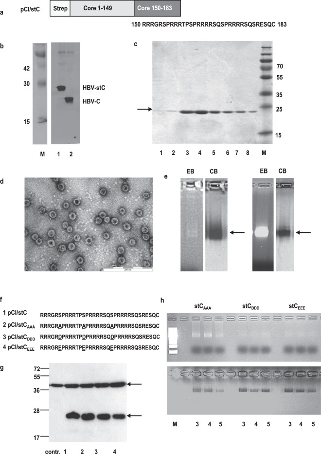
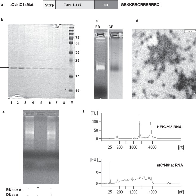
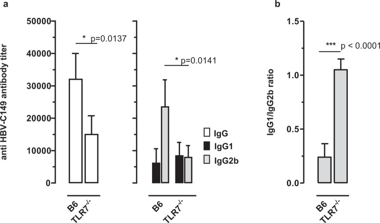
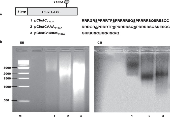
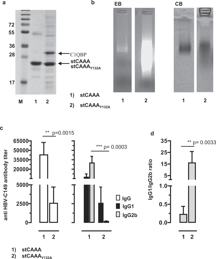
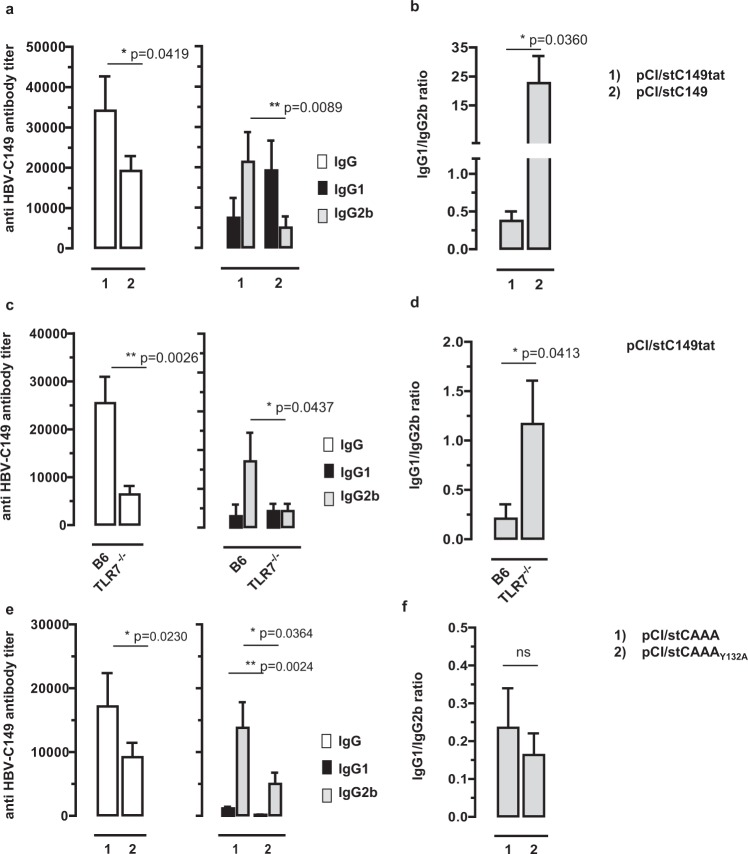
The HBV core protein self-assembles into particles and encapsidates immune-stimulatory bacterial RNA through a cationic COOH-terminal (C150-183) domain. To investigate if different cationic domains have an impact on the endogenous RNA-binding of HBV-C antigens in mammalian cells, we developed a strep-tag (st) based expression/purification system for HBV-C/RNA antigens in vector-transfected HEK-293 cells. We showed that HBV-stC but not HBV-stC149 particles (lacking the cationic domain) capture low amounts of mammalian RNA. Prevention of specific phosphorylation in cationic domains, either by exchanging the serine residues S155, S162 and S170 with alanines (HBV-stCAAA) or by exchanging the entire cationic domain with a HIV-tat48-57-like sequence (HBV-stC149tat) enhanced the encapsidation of RNA into mutant core particles. Particle-bound mammalian RNA functioned as TLR-7 ligand and induced a Th1-biased humoral immunity in B6 but not in TLR-7-/- mice by exogenous (protein) and endogenous (DNA) vaccines. Compared to core particles, binding of mammalian RNA to freely exposed cationic domains in assembly-deficient antigens was enhanced. However, RNA bound to non-particulate antigens unleash its Th1-stimulating adjuvant activity by DNA- but not protein-based vaccination. Mammalian RNAs targeted by an endogenously expressed antigen thus function as a natural adjuvant in the host that facilitates priming of Th1-biased immune responses by DNA-based immunization.


Conflict of interest statement

The authors declare no competing interests.

Figures

Figure 1
Figure 1
Expression and production of HBV core antigens. (a) Map of HBV-stC antigen that contains a NH2-terminal strep- tag sequence. The cationic C150-183 domain is shown. (b) HEK-293 cells were transiently transfected with pCI/stC (lane 1) or pCI/C (lane 2) and labelled with 35S-methionine/cysteine (left panel). Cell lysates were immunoprecipitated with a polyclonal rabbit anti HBV-C serum and processed for SDS-PAGE and fluorography of the gels. The original fluorographies of the gel used to generate this cropped figure are shown in Supplementary Fig. S1. (c) 5 × 108 HEK-293 cells were transiently transfected with the pCI/stC vector and purified as described in the M&M section. 10 μl samples were processed for SDS-PAGE analysis followed by Coomassie Blue staining of the gel. Molecular weight marker (in kDa) is shown. Fractions with the highest antigen content (c; 3 and 4) were pooled, concentrated and processed for electron microscopy (the indicated scale bar represents 200 nm) (d) or for native agarose gel electrophoresis followed by ethidium bromide (EB) and subsequent Coomassie Blue (CB) staining of the gels (e; left panels). Furthermore, HBV-stC particles produced in bacteria were purified as described in Supplementary Information and analyzed on a separate agarose gel (e; right panels). The original gels used to generate this cropped figure are shown in Supplementary Fig. S3. (f) Schematic presentation of mutant cationic domains. The substitution of serine residues at positions S155, S162 and S170 in the wt HBV-stC to alanine HBV-stCAAA, aspartic acid HBV-stCDDD or glutamic acid HBV-stCEEE are indicated. (g) Lysates of HEK-293 cells transiently transfected with control pCI (contr.), pCI/stC (lane 1), pCI/stCAAA (lane 2), pCI/stCDDD (lane 3) or pCI/stCEEE (lane 4) vectors were processed on SDS-PAGE followed by western blot analysis as described in Supplementary Information. The positions of beta-actin (upper panel) and respective HBV core antigens (lower panel) are indicated. (h) The respective antigens were produced in HEK-293 cells and fractions 3 to 5 were processed for native agarose gel electrophoresis followed by ethidium bromide (EB) and subsequent Coomassie Blue (CB)-staining of the gels.
Figure 2
Figure 2
Expression and characterization of a mutant HBV-stC149tat antigen. (a) Schematic presentation of the HBV-stC149tat antigen. This antigen contained the HBV-stC149 sequence COOH-terminally fused with a 14-residue cationic HIV-tat48–57-like sequence. (b–d) 5 × 108 HEK-293 cells were transiently transfected with the pCI/stC149tat vector. The HBV-stC149tat fusion protein was purified from cell lysates using StrepTactin sepharose-packed columns. (b) 10 µl of the elution fractions 1-8 were processed SDS-PAGE analysis followed by Coomassie Blue staining of the gel. Molecular weight marker (in kDa) is shown. (c) Fractions with the highest antigen content were pooled and processed for native agarose gel electrophoresis followed by ethidium bromide (EB) and subsequent Coomassie Blue (CB) staining of the gel. The original gel used to generate this cropped figure is shown in Supplementary Fig. S3a. Purified HBV-stC149tat antigen was further analyzed by electron microscopy (d). The indicated scale bar represents 100 nm. (e) Purified HBV-stC149tat particles were incubated with proteinase K and remained either untreated or treated with RNase A or DNase. Samples were subjected to native agarose gel electrophoresis followed by ethidium bromide (EB)-staining of the gel. (f) The length profile of HEK-293 RNA isolated from non-treated cells (upper panel) or particle-bound RNA (lower panel) was analyzed on a Bioanalyzer 2100.
Figure 3
Figure 3
Induction of HBV core-specific antibodies in mice. (a) B6 mice were immunized with recombinant HEK-293-derived stC149tat or stC149 (n = 4/5). Three weeks post injection serum samples were obtained by tail bleeding and HBV core-specific IgG, IgG1 and IgG2b serum antibody titers were determined by end-point dilution ELISA using bacterial rHBV-C149 particles as detection antigen. Mean specific antibody titers in sera ±SD (a) and the calculated IgG1/IgG2a ratios ±SD (b) of a representative experiment (out of two performed experiments) are shown. The statistical significance of differences in IgG, IgG1 and IgG2b antibody titers between stC149tat- and stC149 immune B6 mice were determined by the unpaired Student’s t-test. (c) B6 mice were immunized with recombinant stC149tat or stC149 proteins. Ten days post immunization spleen cells were stimulated ex vivo for 2 days with the HBV-Core-specific I-Ab-binding C128-140 peptide. The specific IFN-γ release into the cell culture supernatants was determined by ELISA. The statistical significance of differences in IFN-γ levels between stC149- and stC149tat-immune mice (groups 2 and 3) were determined by the unpaired Student’s t-test. (a–c) P values of <0.05 (*) and <0.01 (**) were considered statistically significant.
Figure 4
Figure 4
Induction of HBV core-specific antibodies in B6 and TLR7−/− mice. B6 and TLR-7−/− (n = 4/4) were immunized with recombinant HEK-293-derived stC149tat particles. Serum samples were obtained and analysed as described in the M&M section. Mean specific antibody titers in sera ±SD (a) and the calculated IgG1/IgG2a ratios ±SD (b) of a representative experiment (out of two performed experiments) are shown. The statistical significance of differences in IgG and IgG2b antibody titers between stC149tat immune B6 and TLR7−/− mice was determined by the unpaired Student’s t-test. P values of <0.05 (*) and <0.001 (***) were considered statistically significant.
Figure 5
Figure 5
Expression and characterization of assembly deficient Core antigens. (a) Map of HBV-C antigens that contains a NH2-terminal strep tag sequence, an amino acid substitution from tyrosine to alanine at the position 132, and different COOH-terminal cationic domains: the wt stCY132A, the stCAAAY132A (with additional substitutions of serine residues at positions S155, S162 and S170 to alanines) and stC149tatY132A (substitution of the wt C150-183 domain to a 14-residue HIV-tat like sequence). (b) Antigens were purified from cell lysates after transient transfection of 5 × 108 HEK-293 cells with antigen encoding plasmids as described in the M&M. Identical amounts of recombinant antigens (2,5 µg; calculated for same amounts of monomers determined by SDS-PAGE) were processed for native agarose gel electrophoresis. 1 kb DNA ladder is shown. Agarose gel was stained with ethidium bromide (EB) followed by Coomassie Blue (CB) staining.
Figure 6
Figure 6
Expression and characterization of a mutant HBV-stCAAA and HBV-stCAAAY132A antigen. (a) Samples of purified stCAAA and stCAAAY132A antigens were processed for SDS-PAGE and subsequent Coomassie Blue staining of the gel. The positions of the respective Core antigens and the C1QBP co-precipitating with stCAAAY132A are indicated. The molecular weight marker in kDa is shown. (b) Same amounts of stCAAA and the non-particulate stCAAAY132A (2 µg; calculated for same amounts of monomers determined by SDS-PAGE) were subjected to native agarose gels stained with ethidium bromide (EB) and subsequent with Coomassie Blue (CB). The original gel used to generate this cropped figure is shown in Supplementary Fig. S9. (c) B6 mice were immunized with recombinant HEK-293 derived stCAAA and stCAAAY132A antigens (n = 5/6). Serum samples were obtained 21 days post injection and analyzed for Core-specific IgG, IgG1 and IgG2b antibody titers by end-point dilution ELISA using bacterial rHBV-C149 as detection antigen. Mean specific antibody titers in sera ±SD of a representative experiment (out of two performed experiments) (d) and the calculated IgG1/IgG2a ratios ±SD are shown. (c and d) Statistically significant differences between the group 1 and group 2 were determined using the unpaired student’s t-test. P values of <0.01 (**) and p < 0.001 (***) were considered statistically significant.
Figure 7
Figure 7
Characterization of antibody responses induced by DNA vaccines expressing particulate and non-particulate core antigens. (af) Mice were immunized intradermally with 2 μg particle-coated plasmids with the gene gun (see Supplemental protocols). At d21 mice were boosted with the same vectors. The specific serum Ab responses and isotype profiles (IgG, IgG1, IgG2a) were determined 12 days post boost immunization by end-point dilution ELISA using bacterial rHBV-C149 particles as detection antigen and IgG1/IgG2a ratios were calculated. (a,b) B6 mice (n = 3/4) were immunized with pCI/stC149tat or pCI/stC149 vectors. (c,d) B6 and TLR7−/− mice (n = 3/5) were immunized with pCI/stC149tat. (e,f) B6 mice (n = 5/5) were immunized with pCI/stCAAA or pCI/stCAAAY132A plasmid DNA. Mean specific antibody titers in sera (a,c,e) and the calculated IgG1/IgG2a ratios ±SD (b,d,f) of representative experiments (out of two experiments performed) are shown. Where indicated, the statistical significance of differences in IgG, IgG1 and IgG2b antibody titers was determined by the unpaired Students t-test. P values of < 0.05 (*) and < 0.005 (**) were considered statistically significant.

Similar articles

Cited by

References

    1. Kutzler MA, Weiner DB. DNA vaccines: ready for prime time? Nat Rev Genet. 2008;9:776–788. doi: 10.1038/nrg2432. - DOI - PMC - PubMed
    1. Li L, Petrovsky N. Molecular Adjuvants for DNA Vaccines. Curr Issues Mol Biol. 2017;22:17–40. doi: 10.21775/cimb.022.017. - DOI - PubMed
    1. Kurts C, Robinson BW, Knolle PA. Cross-priming in health and disease. Nat. Rev. Immunol. 2010;10:403–414. doi: 10.1038/nri2780. - DOI - PubMed
    1. Liang F, Lore K. Local innate immune responses in the vaccine adjuvant-injected muscle. Clin Transl Immunology. 2016;5:e74. doi: 10.1038/cti.2016.19. - DOI - PMC - PubMed
    1. Wu J, Chen ZJ. Innate immune sensing and signaling of cytosolic nucleic acids. Annu. Rev. Immunol. 2014;32:461–488. doi: 10.1146/annurev-immunol-032713-120156. - DOI - PubMed

Publication types

MeSH terms