. 2018 Oct 2;9(1):4025.
doi: 10.1038/s41467-018-06156-7.

Megahertz serial crystallography

Max O Wiedorn  1   2   3 Dominik Oberthür  1 Richard Bean  4 Robin Schubert  3   5   6 Nadine Werner  5 Brian Abbey  7 Martin Aepfelbacher  8 Luigi Adriano  9 Aschkan Allahgholi  9 Nasser Al-Qudami  4 Jakob Andreasson  10   11   12 Steve Aplin  1 Salah Awel  1   3 Kartik Ayyer  1 Saša Bajt  9 Imrich Barák  13 Sadia Bari  9 Johan Bielecki  4 Sabine Botha  3   5 Djelloul Boukhelef  4 Wolfgang Brehm  1 Sandor Brockhauser  4   14 Igor Cheviakov  8 Matthew A Coleman  15 Francisco Cruz-Mazo  16 Cyril Danilevski  4 Connie Darmanin  7 R Bruce Doak  17 Martin Domaracky  1 Katerina Dörner  4 Yang Du  1 Hans Fangohr  4   18 Holger Fleckenstein  1 Matthias Frank  15 Petra Fromme  19 Alfonso M Gañán-Calvo  16 Yaroslav Gevorkov  1   20 Klaus Giewekemeyer  4 Helen Mary Ginn  21   22 Heinz Graafsma  9   23 Rita Graceffa  4 Dominic Greiffenberg  24 Lars Gumprecht  1 Peter Göttlicher  9 Janos Hajdu  10   11 Steffen Hauf  4 Michael Heymann  25 Susannah Holmes  7 Daniel A Horke  1   3 Mark S Hunter  26 Siegfried Imlau  1 Alexander Kaukher  4 Yoonhee Kim  4 Alexander Klyuev  9 Juraj Knoška  1   2 Bostjan Kobe  27 Manuela Kuhn  9 Christopher Kupitz  28 Jochen Küpper  1   2   3   29 Janine Mia Lahey-Rudolph  1   30 Torsten Laurus  9 Karoline Le Cong  5 Romain Letrun  4 P Lourdu Xavier  1   31 Luis Maia  4 Filipe R N C Maia  10   32 Valerio Mariani  1 Marc Messerschmidt  4 Markus Metz  1 Davide Mezza  24 Thomas Michelat  4 Grant Mills  4 Diana C F Monteiro  3 Andrew Morgan  1 Kerstin Mühlig  10 Anna Munke  10 Astrid Münnich  4 Julia Nette  3 Keith A Nugent  7 Theresa Nuguid  5 Allen M Orville  22 Suraj Pandey  28 Gisel Pena  1 Pablo Villanueva-Perez  1 Jennifer Poehlsen  9 Gianpietro Previtali  4 Lars Redecke  8   30 Winnie Maria Riekehr  30 Holger Rohde  8 Adam Round  4 Tatiana Safenreiter  1 Iosifina Sarrou  1 Tokushi Sato  1   4 Marius Schmidt  28 Bernd Schmitt  24 Robert Schönherr  30 Joachim Schulz  4 Jonas A Sellberg  33 M Marvin Seibert  10 Carolin Seuring  1   3 Megan L Shelby  15 Robert L Shoeman  17 Marcin Sikorski  4 Alessandro Silenzi  4 Claudiu A Stan  34 Xintian Shi  24 Stephan Stern  1   4 Jola Sztuk-Dambietz  4 Janusz Szuba  4 Aleksandra Tolstikova  1 Martin Trebbin  3   35   36 Ulrich Trunk  9 Patrik Vagovic  1   4 Thomas Ve  37 Britta Weinhausen  4 Thomas A White  1 Krzysztof Wrona  4 Chen Xu  4 Oleksandr Yefanov  1 Nadia Zatsepin  38 Jiaguo Zhang  24 Markus Perbandt  3   5   8 Adrian P Mancuso  4 Christian Betzel  3   5   6 Henry Chapman  39   40   41 Anton Barty  42
Affiliations

Megahertz serial crystallography

Max O Wiedorn et al. Nat Commun. .

Abstract

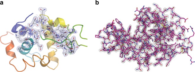
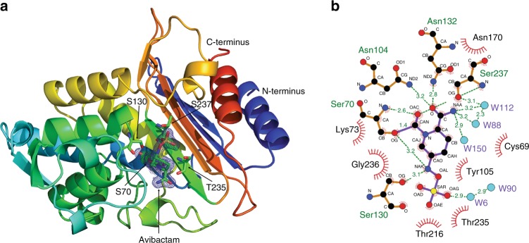
The new European X-ray Free-Electron Laser is the first X-ray free-electron laser capable of delivering X-ray pulses with a megahertz inter-pulse spacing, more than four orders of magnitude higher than previously possible. However, to date, it has been unclear whether it would indeed be possible to measure high-quality diffraction data at megahertz pulse repetition rates. Here, we show that high-quality structures can indeed be obtained using currently available operating conditions at the European XFEL. We present two complete data sets, one from the well-known model system lysozyme and the other from a so far unknown complex of a β-lactamase from K. pneumoniae involved in antibiotic resistance. This result opens up megahertz serial femtosecond crystallography (SFX) as a tool for reliable structure determination, substrate screening and the efficient measurement of the evolution and dynamics of molecular structures using megahertz repetition rate pulses available at this new class of X-ray laser source.


Conflict of interest statement

The authors declare no competing interests.

Figures

Fig. 1
Fig. 1
Megahertz serial crystallography. Pulses from the European XFEL were focused on the interaction region using a set of Beryllium lenses. Protein crystals in crystallization solution were introduced into the focused XFEL beam using a liquid jet of 1.8 µm diameter moving at speeds between 50 m/s and 100 m/s. Diffraction from the sample was measured using an AGIPD, which is capable of measuring up to 3520 pulses per second at megahertz frame rates. In-situ jet imaging (inset) showed that the liquid column does explode under the X-ray illumination conditions of this experiment using a jet with a speed of 100 m/s, but that the liquid jet recovered in less than 1 μs to deliver fresh sample in time for arrival of the next X-ray pulse. Images and movies of jets at different speeds are included in the supplementary material
Fig. 2
Fig. 2
Diffraction pattern from HEWL. Diffraction pattern from a single HEWL microcrystal measured using MHz pulses of 50 fs duration X-rays at 9.3 keV using the AGIPD 1M detector in the SPB/SFX instrument. Dynamic gain switching of the AGIPD detector enables simultaneous low noise and high dynamic range: each pixel has three gain settings which are automatically selected depending on the per-pixel cumulative intensity to simultaneously maximize sensitivity and dynamic range. Image clipped at 2600 counts to show content, full dynamic range of brightest spots extends to 109,000 counts
Fig. 3
Fig. 3
Images of interaction of the EuXFEL liquid jet for the first 5 pulses in a train. Jets in the range of 50–100 m/s recover in time for the next pulse (first three rows), whereas slower jets of the type commonly used at LCLS do not recover in time for the next XFEL pulse at MHz repetition rates (bottom row). The bottom line provides linkage back to the results presented in ref. . Red line shows the intersection point with X-ray pulses. Images obtained by synchronized laser back illumination. Movies with finer time steps are included as supplementary material
Fig. 4
Fig. 4
HEWL diffraction was measured on all pulses in the pulse train. a Hit fraction as a function of pulse number indicates that crystals are hit randomly on any pulse within the MHz EuXFEL pulse train, and not only on the first pulse in the pulse train. b Indexable lattices were equally distributed among the MHz XFEL pulse trains and no sign of degradation in data quality is observed through the pulse train as measured by the overall CC* for subsets of the data corresponding to each pulse. c CrystFEL resolution estimate as a function of X-ray pulse within a train shows no decrease in estimated resolution through the course of the pulse train. d CC* for data separated from each pulse indicates similar data quality for each pulse in the pulse train. Merging all pulses produces higher data quality (as expected). e Correlation of merged data from the first pulse relative to each subsequent pulse in the pulse train indicates that data are similar on each pulse to the limit of data quality available in this experiment. Both d and e are generated from the same stream files used for structure determination sorted according to pulse ID
Fig. 5
Fig. 5
Electron density map for HEWL by MHz SFX. a 2Fo-Fc map at 1 sigma overlaid on Fo-Fc map at 3 sigma from molecular replacement using a solvent-free version of the 4ET8 lysozyme structure as the starting model. b Integrity of the measured data is verified by complete rebuilding of the structure from a truncated starting model after removal of residues 1–16 and 40–60 of the polypeptide chain using Autobuild
Fig. 6
Fig. 6
Structure of CTX-M-14 β-lactamase determined by MHz SFX. a 2Fo-Fc map at 1 sigma overlaid on Fo-Fc map at 3 sigma around covalently bound avibactam from molecular replacement using a solvent-free version of the 5TWD β-lactamase structure from ref. as the starting model. b Representation of covalently bound avibactam to OG of Ser70, stabilized by hydrogen bonds and hydrophobic interactions with surrounding amino acids of CTX-M-14. Figure was prepared using Ligplot

References

    1. Chapman HN, et al. Femtosecond X-ray protein nanocrystallography. Nature. 2011;470:73–77. doi: 10.1038/nature09750. - DOI - PMC - PubMed
    1. Boutet S, et al. High-resolution protein structure determination by serial femtosecond crystallography. Science. 2012;337:362–364. doi: 10.1126/science.1217737. - DOI - PMC - PubMed
    1. Kang Y, et al. Crystal structure of rhodopsin bound to arrestin by femtosecond X-ray laser. Nature. 2015;523:561–567. doi: 10.1038/nature14656. - DOI - PMC - PubMed
    1. Pande K, et al. Femtosecond structural dynamics drives the trans/cis isomerization in photoactive yellow protein. Science. 2016;352:725–729. doi: 10.1126/science.aad5081. - DOI - PMC - PubMed
    1. Stagno JR, et al. Structures of riboswitch RNA reaction states by mix-and-inject XFEL serial crystallography. Nature. 2017;541:242–246. doi: 10.1038/nature20599. - DOI - PMC - PubMed

Publication types