. 2018 Oct 2;8(1):14683.
doi: 10.1038/s41598-018-33048-z.

How phosphorylation influences E1 subunit pyruvate dehydrogenase: A computational study

Affiliations

How phosphorylation influences E1 subunit pyruvate dehydrogenase: A computational study

Jacopo Sgrignani et al. Sci Rep. .

Abstract

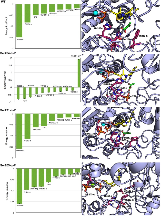
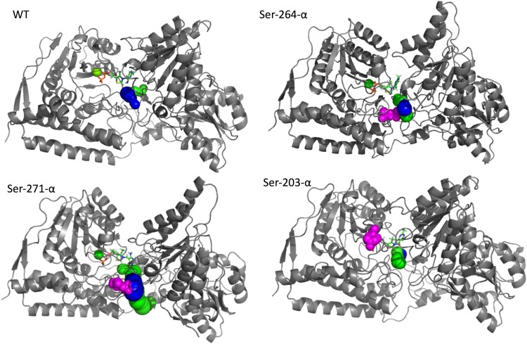
Pyruvate (PYR) dehydrogenase complex (PDC) is an enzymatic system that plays a crucial role in cellular metabolism as it controls the entry of carbon into the Krebs cycle. From a structural point of view, PDC is formed by three different subunits (E1, E2 and E3) capable of catalyzing the three reaction steps necessary for the full conversion of pyruvate to acetyl-CoA. Recent investigations pointed out the crucial role of this enzyme in the replication and survival of specific cancer cell lines, renewing the interest of the scientific community. Here, we report the results of our molecular dynamics studies on the mechanism by which posttranslational modifications, in particular the phosphorylation of three serine residues (Ser-264-α, Ser-271-α, and Ser-203-α), influence the enzymatic function of the protein. Our results support the hypothesis that the phosphorylation of Ser-264-α and Ser-271-α leads to (1) a perturbation of the catalytic site structure and dynamics and, especially in the case of Ser-264-α, to (2) a reduction in the affinity of E1 for the substrate. Additionally, an analysis of the channels connecting the external environment with the catalytic site indicates that the inhibitory effect should not be due to the occlusion of the access/egress pathways to/from the active site.


Conflict of interest statement

The authors declare no competing interests.

Figures

Figure 1
Figure 1
(I) Schematic view of the three PDC subunits with the corresponding reaction schemes. E1 (pink, pyruvate dehydrogenase) catalyzes pyruvate decarboxylation. E2 (blue, dihydrolipoyl trans-acetylase) catalyzes the acetyl transfer to CoA. E3 (white, dihydrolipoyl dehydrogenase) catalyzes the regeneration of the lipoamide moiety. (II) General PDC catalyzed reaction.
Figure 2
Figure 2
Differences between the RMSF values measured for the three phosphorylated species, Ser-264-α-P (light blue), Ser-271-α-P (orange), Ser-203-α-P (violet), and the WT. Positive values correspond to an increment of RMSF values with respect to WT, while negative values correspond to a reduction. The data for the α (A) and β (B) subunits are reported in separate diagrams for clarity.
Figure 3
Figure 3
(A) Backbone RMSD calculated with respect to the starting coordinates of the WT protein over the entire MD simulations. WT (violet) and the three phosphorylated species, Ser-264-α-P (light blue), Ser-271-α-P (orange), and Ser-203-α-P (green)). (B) Binding site RMSD (His63-α, Met200-α, His263-α, Phe61-α, Tyr89-α, Met82-β) calculated for the entire residues (backbone + sidechains) over the entire simulation time. WT (violet) and the three phosphorylated species Ser-264-α-P (light blue), Ser-271-α-P (orange), and Ser-203-α-P (green).
Figure 4
Figure 4
PDC-E1 structure colored by calculated B-factor (computed with the RMSF tool implemented in GROMACS 5.1.4). (A) Wild type, (B) Ser-264-α-P, (C) Ser-271-α-P, (D) Ser-203-α-P.
Figure 5
Figure 5
(Left column), the results of per-residue Energy decomposition carried out for the WT (A) and the systems bearing a Ser-264-α-P, Ser-271-α-P, or Ser-203-α-P. For sake of clarity, only contributions higher the 0.2 kcal/mol (absolute value) were reported. (Right column) representative structure of the catalytic site in the four investigated systems. The residues with the higher contributions to the binding and the phosphorylated residues are depicted as sticks; the nonpolar hydrogens are not shown for the sake of clarity.
Figure 6
Figure 6
Graphical representation of the Caver3.0 results. The first and the second tunnels are represented in blue and green, respectively. The three phosphorylated serine residues are always depicted in magenta as spatial references. In the cases of Ser-264-α-P and Ser-271-α-P, in which the tunnels have a common pathway, only one color is used to depict the common part.

References

    1. Patel MS, Nemeria NS, Furey W, Jordan F. The pyruvate dehydrogenase complexes: structure-based function and regulation. J Biol Chem. 2014;289:16615–16623. doi: 10.1074/jbc.R114.563148. - DOI - PMC - PubMed
    1. Solaini G, Sgarbi G, Baracca A. Oxidative phosphorylation in cancer cells. Biochimica et Biophysica Acta (BBA) - Bioenergetics. 2011;1807:534–542. doi: 10.1016/j.bbabio.2010.09.003. - DOI - PubMed
    1. Ahlqvist KJ, Suomalainen A, Hämäläinen RH. Stem cells, mitochondria and aging. Biochimica et Biophysica Acta (BBA) - Bioenergetics. 2015;1847:1380–1386. doi: 10.1016/j.bbabio.2015.05.014. - DOI - PubMed
    1. He H, Xia H, Xia Q, Ren Y, He H. Design and optimization of N-acylhydrazone pyrimidine derivatives as E. coli PDHc E1 inhibitors: Structure-activity relationship analysis, biological evaluation and molecular docking study. Bioorg. Med. Chem. 2017;25:5652-. doi: 10.1016/j.bmc.2017.08.038. - DOI - PubMed
    1. Zhou Y, et al. Design, Synthesis, and Potency of Pyruvate Dehydrogenase Complex E1 Inhibitors against Cyanobacteria. Biochemistry. 2017;56:6491–6502. doi: 10.1021/acs.biochem.7b00636. - DOI - PubMed

Publication types

MeSH terms

Substances