. 2018 Oct 2;8(1):14652.
doi: 10.1038/s41598-018-32376-4.

Simvastatin Protects Cardiomyocytes Against Endotoxin-induced Apoptosis and Up-regulates Survivin/NF-κB/p65 Expression

Affiliations

Simvastatin Protects Cardiomyocytes Against Endotoxin-induced Apoptosis and Up-regulates Survivin/NF-κB/p65 Expression

Lana Nežić et al. Sci Rep. .

Abstract

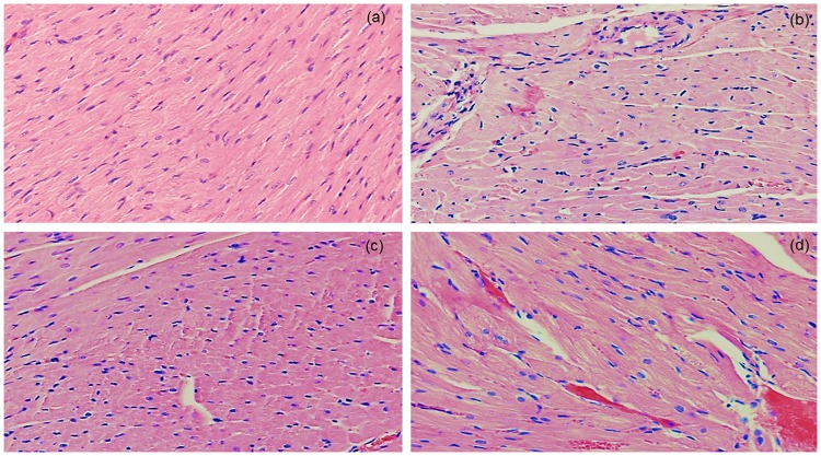
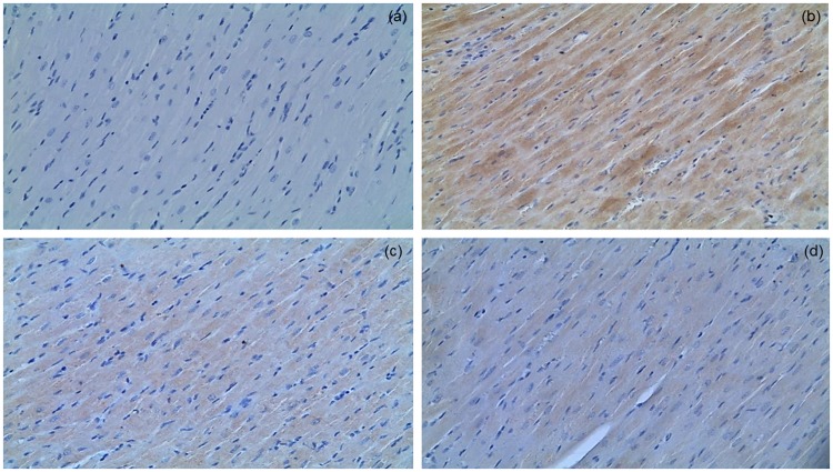
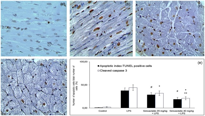
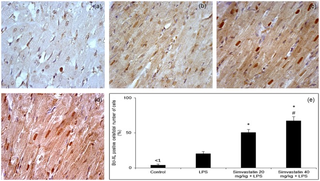
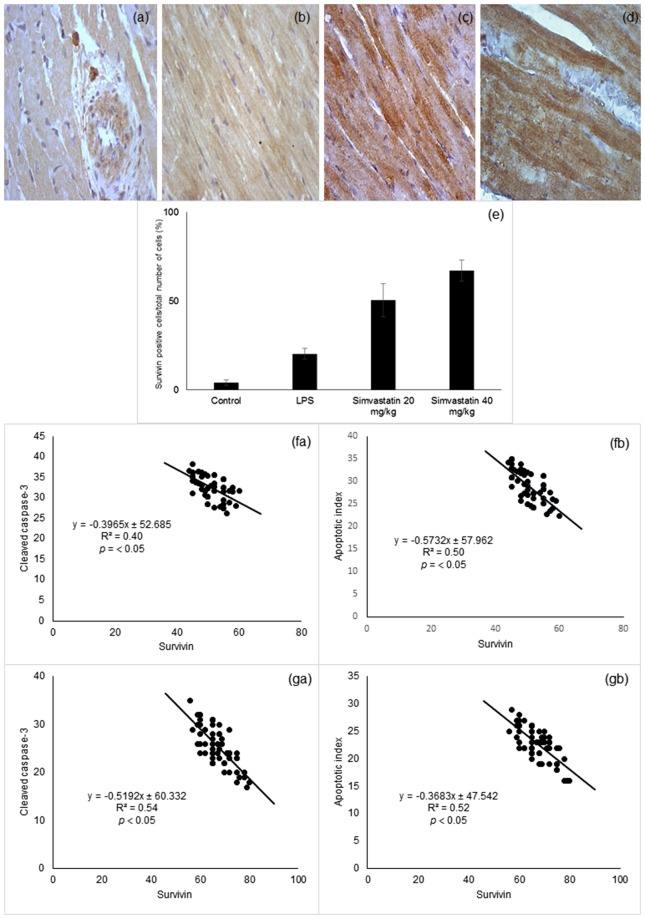
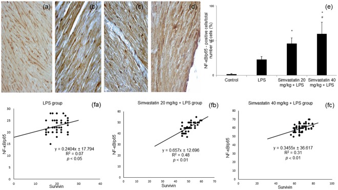
This study is aimed to investigate whether simvastatin induces cardiomyocytes survival signaling in endotoxin (lipopolysaccharide, LSP)-induced myocardial injury, and if so, further to determine a role of survivin in simvastatin-anti-apoptotic effect. Wistar rats were pretreated with simvastatin (10-40 mg/kg po) before a single non-lethal dose of LPS. In myocardial tissue, LPS induced structural disorganization of myofibrils with significant inflammatory infiltrate (cardiac damage score, CDS = 3.87 ± 0.51, p < 0.05), whereas simvastatin dose-dependently abolished structural changes induced by LPS (p < 0.01). Simvastatin in 20 mg/kg and 40 mg/kg pretreatment, dose dependently, attenuated myocardial apoptosis determined as apoptotic index (28.8 ± 4.5% and 18.9 ± 3.5, p < 0.05), decreased cleaved caspase-3 expression (32.1 ± 5.8%, p < 0.01), along with significant Bcl-xL expression in the simvastatin groups (p < 0.01). Interestingly, in the simvastatin groups were determined significantly increased expression of survivin (p < 0.01), but in negative correlation with cleaved caspase-3 and apoptotic indices (p < 0.01). Simvastatin has a cardioprotective effects against LPS induced apoptosis. The effect may be mediated by up-regulation of survivin via activation of NF-κB, which leads to reduced activation of caspase-3 and consequent apoptosis of cardiomyocytes in experimental sepsis.


Conflict of interest statement

The authors declare no competing interests.

Figures

Figure 1
Figure 1
Pretreatment with simvastatin protected from LPS-induced myocardial injury. Light micrographs of the myocardial tissue, H&E, magnifications 100x and 200x). (a) Control group showed normal histology. (b) LPS group. (c) Simvastatin 20 group + LPS. (d) Simvastatin 40 group + LPS. Note decreased severe myocardial lesions and maintenance of the normal histology of the simvastatin-treated myocardium in contrast to the deterioration of myocardial tissue in the LPS-challenged group.
Figure 2
Figure 2
Simvastatin inhibited apoptosis in rat myocardial tissue in LPS induced inflammation by down-regulating cleaved caspase 3 expression. Representative images with quantitative analysis of apoptotic cardiomyocytes that were challenged with LPS for induction of inflammation and either pretreated with simvastatin 20 and simvastatin 40 before LPS. The expression of cleaved caspase 3 in rat myocardial tissue examined by immunohistochemical staining, magnification 200x. (a) Control group. (b) Intense cytoplasmic staining of cleaved caspase 3 in cardiomyocytes in LPS group, as characteristic of cell in apoptosis. Marked reduction of apoptotic cardiomyocytes in the simvastatin 20 (c) and simvastatin 40 (d) groups.
Figure 3
Figure 3
Simvastatin inhibited apoptosis in rat myocardial tissue in LPS induced inflammation detected by TUNEL staining, magnification 400x. Brown stained nuclei indicate TUNEL-positive cardiomyocytes. The apoptosis increased significantly in the LPS (b) and simvastatin group (c) compared with the control group (a). Note that induction of sepsis by LPS resulted in a marked appearance of TUNEL-positive cardiomyocytes (arrow) quantified and shown as AIs (black columns) (e), which was significantly reduced by simvastatin 20 (c) and simvastatin 40 (D). (e) Quantitative analysis of apoptotic cells counted in immunohistochemically stained myocardial sections for cleaved caspase 3 and corresponding frequencies of TUNEL positive cardiomyocytes are shown, *p < 0.01 in comparation with LPS group, **p < 0.05 in comparation with simvastatin 20, #p < 0.05 in comparation with LPS group.
Figure 4
Figure 4
Simvastatin increased Bcl-XL expression in rat myocardial tissue in LPS induced inflammation. Representative images with quantitative analysis of distribution of Bcl-XL staining in cardiomyocytes that were challenged with LPS for induction of inflammation or either pretreated with simvastatin 20 and simvastatin 40 before LPS. The expression of Bcl-XLin rat myocardial tissue examined by immunohistochemical staining, magnification 400x. (a) Control group. (b) Note focally distributed immunopositive BCL-XL cardiomyocytes in the LPS group. In the simvastatin group 20 (c) and simvastatin 40 (d) BCL-XL expression was significantly intensive and widely distributed in cardiomyocytes. (e) Quantitative analysis of distribution of Bcl-XL immune-positivity cells in selected fields, *p < 0.01 in comparation with LPS group, #p < 0.01 in comparation with simvastatin 20 group.
Figure 5
Figure 5
Simvastatin increased survivin expression in rat myocardial tissue in LPS induced inflammation. Representative images with semi-quantitative analysis of survivin positive cells in myocardium in groups that were challenged with LPS or either pretreated with simvastatin 20 or simvastatin 40 before LPS. Survivin expression in rat myocardial tissue examined by immunohistochemical staining, magnification 400x. (a) Control group. (b) LPS group. Note intensive survivin cytoplasmic staining of cardiomyocytes in the simvastatin 20 (c) and simvastatin 40 (d). (e) Semiquantitative analysis of survivin expression, *p < 0.01 in comparation with LPS group, #p < 0.01 in comparation with simvastatin 20 group. (f) The correlation of survivin expression and cardiomyocytes apoptosis determined by cleaved-caspase-3 (fa) and apoptotic index (fb) in the group pretreated with simvastatin 20. (g) The correlation of survivin expression and cardiomyocytes apoptosis determined by cleaved-caspase-3 (ga) and apoptotic index (gb) in the group pretreated with simvastatin 40.
Figure 6
Figure 6
Simvastatin increased NF-κB expression in rat myocardial tissue in LPS induced inflammation. Representative images with semi-quantitative analysis of survivin positive cells in myocardium in groups that were challenged with LPS or either pretreated with simvastatin 20 or simvastatin 40 before LPS. Immunohistological staining of myocardial tissues was performed using a p65-specific antibody to evaluate NF-κB p65 expression in cardiomyocytes, magnification 200x and 400x. (a) Control group. Note subsets of cardiomyocytes positive for NF-κB/p65 in the cell cytoplasm and/or nucleus in the LPS group (b), and intensive nuclear immunostaining in the simvastatin 20 (c) and simvastatin 40 (d) groups. (e) Semiquantitative analysis of NF-κB/p65 expression. *p < 0.01 in comparation with LPS group, #p < 0.05 in comparation with simvastatin 20 group. (f) The correlations of survivin expression and NF-κB/p65 positive cardiomyocytes within the experimental groups (fa, fb, fc).

References

    1. Li Y, Ge S, Peng Y. Inflammation and cardiac dysfunction during sepsis, muscular dystrophy, and myocarditis. Burns Trauma. 2013;1:109–121. doi: 10.4103/2321-3868.123072. - DOI - PMC - PubMed
    1. Romero-Bermejo FJ, et al. Sepsis-induced cardiomyopathy. Curr. Cardiol. Rev. 2011;7:163–183. doi: 10.2174/157340311798220494. - DOI - PMC - PubMed
    1. Hobai IA, et al. Dysregulation of intracellular calcium transporters in animal models of sepsis induced cardiomyopathy. Shock. 2015;43:3–15. doi: 10.1097/SHK.0000000000000261. - DOI - PMC - PubMed
    1. Cimolai MC, et al. Mitochondrial Mechanisms in Septic Cardiomyopathy. Int. J. Mol. Sci. 2015;16:17763–17778. doi: 10.3390/ijms160817763. - DOI - PMC - PubMed
    1. Smeding L, et al. Structural changes of the heart during severe sepsis or septic shock. Shock. 2012;37:449–456. doi: 10.1097/SHK.0b013e31824c3238. - DOI - PubMed

Publication types

MeSH terms