. 2018 Sep 28:155:32.
doi: 10.1186/s41065-018-0069-1. eCollection 2018.

Long-read sequencing identified a causal structural variant in an exome-negative case and enabled preimplantation genetic diagnosis

Affiliations

Long-read sequencing identified a causal structural variant in an exome-negative case and enabled preimplantation genetic diagnosis

Hefan Miao et al. Hereditas. .

Abstract

Background: For a proportion of individuals judged clinically to have a recessive Mendelian disease, only one heterozygous pathogenic variant can be found from clinical whole exome sequencing (WES), posing a challenge to genetic diagnosis and genetic counseling. One possible reason is the limited ability to detect disease causal structural variants (SVs) from short reads sequencing technologies. Long reads sequencing can produce longer reads (typically 1000 bp or longer), therefore offering greatly improved ability to detect SVs that may be missed by short-read sequencing.

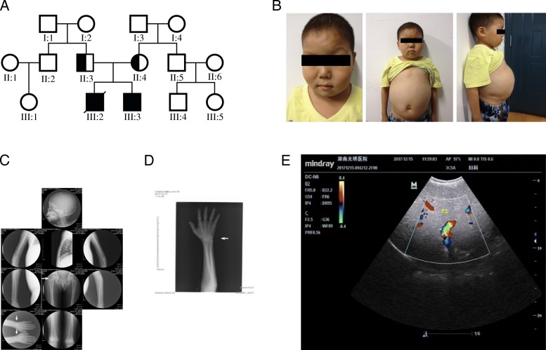
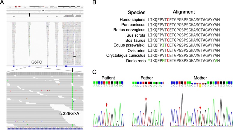
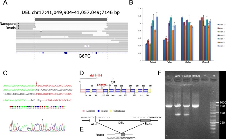
Results: Here we describe a case study, where WES identified only one heterozygous pathogenic variant for an individual suspected to have glycogen storage disease type Ia (GSD-Ia), which is an autosomal recessive disease caused by bi-allelic mutations in the G6PC gene. Through Nanopore long-read whole-genome sequencing, we identified a 7.1 kb deletion covering two exons on the other allele, suggesting that complex structural variants (SVs) may explain a fraction of cases when the second pathogenic allele is missing from WES on recessive diseases. Both breakpoints of the deletion are within Alu elements, and we designed Sanger sequencing and quantitative PCR assays based on the breakpoints for preimplantation genetic diagnosis (PGD) for the family planning on another child. Four embryos were obtained after in vitro fertilization (IVF), and an embryo without deletion in G6PC was transplanted after PGD and was confirmed by prenatal diagnosis, postnatal diagnosis, and subsequent lack of disease symptoms after birth.

Conclusions: In summary, we present one of the first examples of using long-read sequencing to identify causal yet complex SVs in exome-negative patients, which subsequently enabled successful personalized PGD.

Keywords: G6PC; GSD-Ia; Long-read sequencing; PGD; Structural variants; WES; Whole-exome sequencing.


Conflict of interest statement

The research including human subjects, human material, human data, has been performed in accordance with the Declaration of Helsinki and was approved by the ethics committee of Reproductive and Genetic Hospital of CITIC-Xiangya.The research contains individual person’s data (including detailed clinical phenotypes and images) and we have obtained consent from parents of the children.J.Z., Q.Y., F.L., D.W. are employees and K.W. is consultant of Grandomics Biosciences. All the other authors have no competing interests.Springer Nature remains neutral with regard to jurisdictional claims in published maps and institutional affiliations.

Figures

Fig. 1
Fig. 1
Clinical characteristics of the proband. (a) Pedigree of the family. III:3 represents the proband, whose older brother (III:2) has decreased. (b) The clinical features include a rounded doll’s face, fatty cheeks and protuberant abdomen. (c) X-ray films of the whole body of the patient. White arrows mark areas with obvious osteoporosis. (d) Focused view of X-ray film on the hand of the proband, where the wrist marked by white arrows has obvious osteoporosis. (e) Image of type-B ultrasonic on the proband shows severe liver enlargement. Blue color: The blood flow away from the detector of ultrasound B-mode scanner; Red color: The blood flow to the detector of ultrasound B-mode scanner
Fig. 2
Fig. 2
Identification of a c.326G > A missense mutation in the G6PC gene. (a) Whole-exome sequencing identified a homozygous c.326G > A missense variant in exon 2 of the G6PC gene. (b) The amino acid 109 (marked by red color) affected by c.326G > A is highly conserved across different species. (c) Sanger sequencing on the pedigree showed that the father does not carry the c.326G > A missense variant and that the mother carries a heterozygous c.326G > A missense variant
Fig. 3
Fig. 3
Long-read sequencing identified a deletion in the G6PC gene. (a) IGV screen shot of reads at the G6PC locus. Four reads carry a deletion (chr17 g.41049904_41057049del7125 that starts from the first intron of the LINC00671 gene to intron 2 of the G6PC gene. (b) Quantitative PCR validation of the deletion in the trio. Relative quantitation (RQ) of copy number was analyzed by the ΔΔCT method, and error bars represent standard deviation. The deletion includes exon 1F (5′-Flanking introns), exon 1 and exon 2, and the patient and his father are mutation carriers while his mother is normal. (c) Sanger validation of the deletion breakpoints. The first sequence shows the mutated genomic segment, while the second and third sequences show expected genomic segments if deletion is not present. The red arrow refers to the breakpoint, and a 7125 bp sequence is deleted based on the human reference genome (GRCh37). (d) Depiction of the protein domains that were targeted by the non-synonymous mutation and the 7.1 kb deletion. (e) Illustration of the genomic contexts of the two breakpoints, which are both located in known Alu elements. (f) Gel electrophoresis of the PCR product designed to detect the deletion. The lane marked with M represent GeneRuler 50 bp DNA Ladder (Thermo Scientific™), and all lanes (except “-“lane) include an ~ 800 bp internal control (β-Globin gene). A 418 bp fragment can be amplified from the father and the proband

References

    1. Posey JE, Rosenfeld JA, James RA, Bainbridge M, Niu Z, Wang X, Dhar S, Wiszniewski W, Akdemir ZH, Gambin T, et al. Molecular diagnostic experience of whole-exome sequencing in adult patients. Genet Med. 2016;18(7):678–685. doi: 10.1038/gim.2015.142. - DOI - PMC - PubMed
    1. Yang Y, Muzny DM, Reid JG, Bainbridge MN, Willis A, Ward PA, Braxton A, Beuten J, Xia F, Niu Z, et al. Clinical whole-exome sequencing for the diagnosis of mendelian disorders. N Engl J Med. 2013;369(16):1502–1511. doi: 10.1056/NEJMoa1306555. - DOI - PMC - PubMed
    1. Retterer K, Juusola J, Cho MT, Vitazka P, Millan F, Gibellini F, Vertino-Bell A, Smaoui N, Neidich J, Monaghan KG, et al. Clinical application of whole-exome sequencing across clinical indications. Genet Med. 2016;18(7):696–704. doi: 10.1038/gim.2015.148. - DOI - PubMed
    1. Trujillano D, Bertoli-Avella AM, Kumar Kandaswamy K, Weiss ME, Koster J, Marais A, Paknia O, Schroder R, Garcia-Aznar JM, Werber M, et al. Clinical exome sequencing: results from 2819 samples reflecting 1000 families. Eur J Hum Genet. 2017;25(2):176–182. doi: 10.1038/ejhg.2016.146. - DOI - PMC - PubMed
    1. Lee H, Deignan JL, Dorrani N, Strom SP, Kantarci S, Quintero-Rivera F, Das K, Toy T, Harry B, Yourshaw M, et al. Clinical exome sequencing for genetic identification of rare Mendelian disorders. Jama. 2014;312(18):1880–1887. doi: 10.1001/jama.2014.14604. - DOI - PMC - PubMed

LinkOut - more resources