

Assessment of myocardial fibrosis using T1-mapping and extracellular volume measurement on cardiac magnetic resonance imaging for the diagnosis of radiation-induced cardiomyopathy
- PMID: 30279930
- PMCID: PMC6149610
- DOI: 10.1016/j.jccase.2018.06.001
Assessment of myocardial fibrosis using T1-mapping and extracellular volume measurement on cardiac magnetic resonance imaging for the diagnosis of radiation-induced cardiomyopathy
Abstract
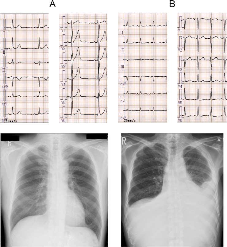
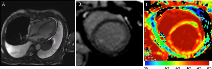
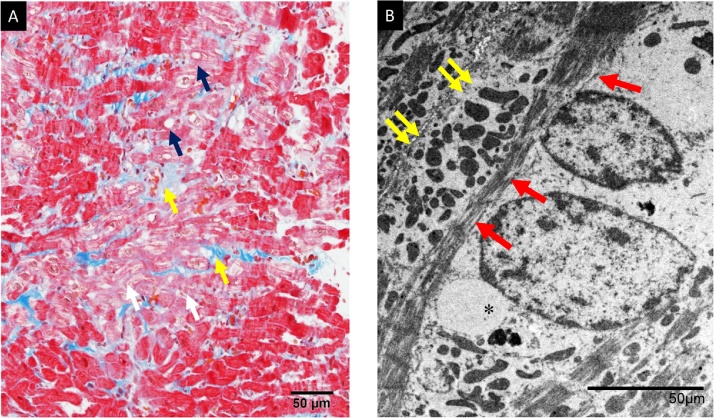
Radiation-induced heart disease (RIHD) is a serious side effect of thoracic radiation therapy (RT) and is associated with significant morbidity and mortality. Radiation-induced cardiomyopathy (RICM) is one of the manifestations of RIHD, which represents with left ventricular (LV) systolic and diastolic dysfunction due to myocardial fibrosis. Although the diagnosis of RIHD is challenging and is generally an exclusion diagnosis, multimodality imaging including echocardiography, cardiac computed tomography and cardiac magnetic resonance (CMR) imaging could help the diagnosis. Herein, we report a case of 70-years-old male, who had been treated with chemo-radiation therapy for early esophageal cancer, was suffered from medically refractory heart failure due to severely reduced LV systolic function and constrictive pericarditis 8 years after chemo-radiation therapy. Although no gadolinium-enhancement (LGE) was detected on CMR, T1 mapping depicted increased extracellular matrix volumes of 45%, which suggested global myocardial fibrosis. Histopathological analysis by endomyocardial biopsy (EBM) revealed marked degeneration of myocytes and interstitial fibrosis, while vacuolation in myocytes which is characteristics of chemotherapy induced cardiomyopathy was not specific by electron microscopy. Therefore, we diagnosed that the present case was likely to the RICM. <Learning objective: RICM is characterized by inflammation followed by the development of a diffuse, patchy interstitial fibrosis of the myocardium, which is usually obtained either by EBM or at autopsy. Native and post-contrast T1-mapping by CMR enables to estimate extracellular volume (ECV), which is believed to be increased as a result of diffuse myocardial fibrosis. The assessment of myocardial fibrosis using ECV should be useful for early detection of myocardial damage due to RT, and which probably taking place of EBM.>.
Keywords: Myocardial fibrosis; Onco-cardiology; Radiation-induced heart disease.
Figures
References
-
- Lipshultz S.E., Adams M.J. Cardiotoxicity after childhood cancer: beginning with the end in mind. J Clin Oncol. 2010;28:1276–1281. - PubMed
-
- Yeh E.T., Bickford C.L. Cardiovascular complications of cancer therapy: incidence, pathogenesis, diagnosis, and management. J Am Coll Cardiol. 2009;53:2231–2247. - PubMed
-
- Lancellotti P., Nkomo V.T., Badano L.P., Bergler-Klein J., Bogaert J., Davin L. Expert consensus for multi-modality imaging evaluation of cardiovascular complications of radiotherapy in adults: a report from the European Association of Cardiovascular Imaging and the American Society of Echocardiography. Eur Heart J Cardiovasc Imaging. 2013;14:721–740. - PubMed
-
- Kim R.J., Wu E., Rafael A., Chen E.L., Parker M.A., Simonetti O. The use of contrast-enhanced magnetic resonance imaging to identify reversible myocardial dysfunction. N Engl J Med. 2000;343:1445–1453. - PubMed
-
- Umezawa R., Ota H., Takanami K., Ichinose A., Matsushita H., Saito H. MRI findings of radiation-induced myocardial damage in patients with oesophageal cancer. Clin Radiol. 2014;69:1273–1279. - PubMed
Publication types
LinkOut - more resources
Full Text Sources
