

Dicer deficiency in proximal tubules exacerbates renal injury and tubulointerstitial fibrosis and upregulates Smad2/3
- PMID: 30280598
- PMCID: PMC6337000
- DOI: 10.1152/ajprenal.00402.2018
Dicer deficiency in proximal tubules exacerbates renal injury and tubulointerstitial fibrosis and upregulates Smad2/3
Abstract
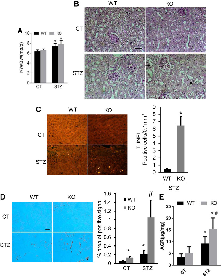
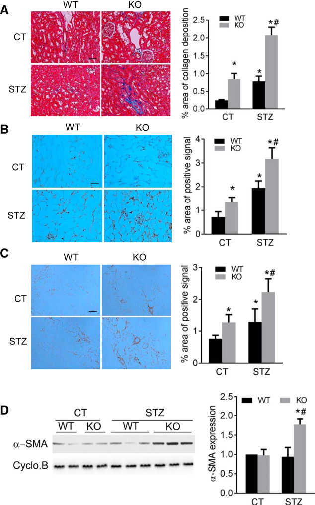
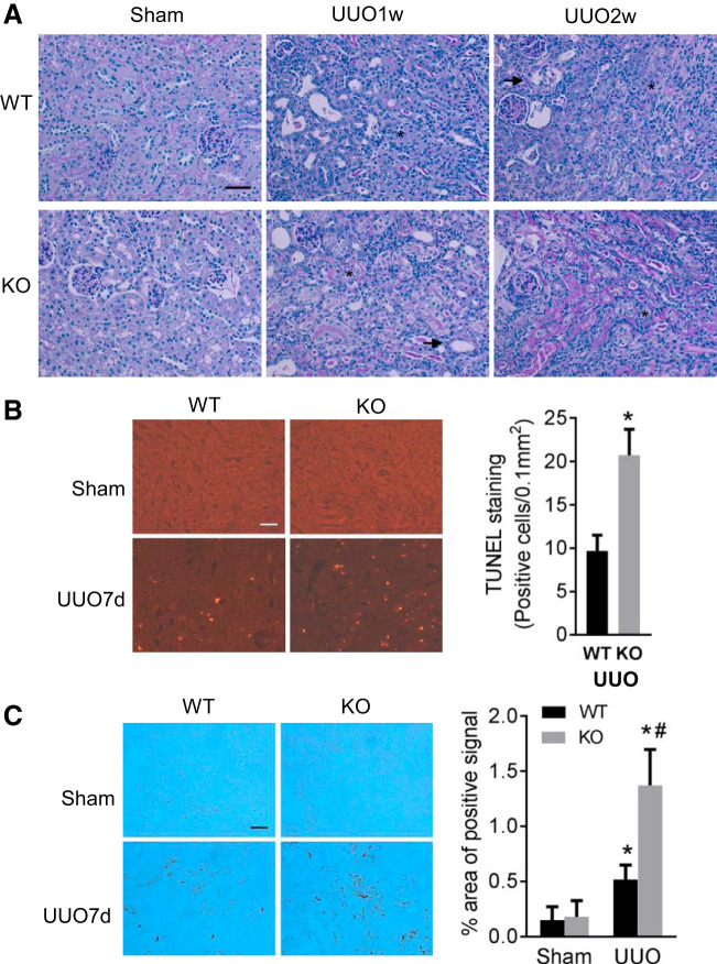
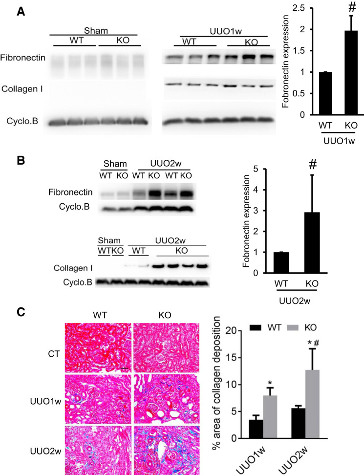
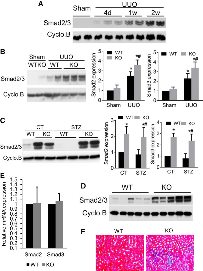
Renal fibrosis is a common pathological feature in chronic kidney disease (CKD), including diabetic kidney disease (DKD) and obstructive nephropathy. Multiple microRNAs have been implicated in the pathogenesis of both DKD and obstructive nephropathy, although the overall role of microRNAs in tubular injury and renal fibrosis in CKD is unclear. Dicer (a key RNase III enzyme for microRNA biogenesis) was specifically ablated from kidney proximal tubules in mice via the Cre-lox system to deplete micoRNAs. Proximal tubular Dicer knockout (PT- Dicer KO) mice and wild-type (WT) littermates were subjected to streptozotocin (STZ) treatment to induce DKD or unilateral ureteral obstruction (UUO) to induce obstructive nephropathy. Renal hypertrophy, renal tubular apoptosis, kidney inflammation, and tubulointerstitial fibrosis were examined. Compared with WT mice, PT- Dicer KO mice showed more severe tubular injury and renal inflammation following STZ treatment. These mice also developed higher levels of tubolointerstitial fibrosis. Meanwhile, PT- Dicer KO mice had a significantly higher Smad2/3 expression in kidneys than WT mice (at 6 mo of age) in both control and STZ-treated mice. Similarly, UUO induced more severe renal injury, inflammation, and interstitial fibrosis in PT- Dicer KO mice than WT. Although we did not detect obvious Smad2/3 expression in sham-operated mice (2-3 mo old), significantly more Smad2/3 was induced in obstructed PT- Dicer KO kidneys. These results supported a protective role of Dicer-dependent microRNA synthesis in renal injury and fibrosis development in CKD, specifically in DKD and obstructive nephropathy. Depletion of Dicer and microRNAs may upregulate Smad2/3-related signaling pathway to enhance the progression of CKD.
Keywords: Dicer; Smad2/3; diabetic kidney disease; fibrosis; microRNA; unilateral urethral obstruction.
Conflict of interest statement
No conflicts of interest, financial or otherwise, are declared by the authors.
Figures
Similar articles
-
Loss of angiotensin-converting enzyme 2 enhances TGF-β/Smad-mediated renal fibrosis and NF-κB-driven renal inflammation in a mouse model of obstructive nephropathy.Lab Invest. 2012 May;92(5):650-61. doi: 10.1038/labinvest.2012.2. Epub 2012 Feb 13. Lab Invest. 2012. PMID: 22330342
-
Tubular NOX4 expression decreases in chronic kidney disease but does not modify fibrosis evolution.Redox Biol. 2019 Sep;26:101234. doi: 10.1016/j.redox.2019.101234. Epub 2019 Jun 5. Redox Biol. 2019. PMID: 31247506 Free PMC article.
-
MicroRNA-214 antagonism protects against renal fibrosis.J Am Soc Nephrol. 2014 Jan;25(1):65-80. doi: 10.1681/ASN.2013010072. Epub 2013 Oct 24. J Am Soc Nephrol. 2014. PMID: 24158985 Free PMC article.
-
Obstructive nephropathy and molecular pathophysiology of renal interstitial fibrosis.Physiol Rev. 2023 Oct 1;103(4):2827-2872. doi: 10.1152/physrev.00027.2022. Epub 2023 Jul 13. Physiol Rev. 2023. PMID: 37440209 Free PMC article. Review.
-
The proximal tubule in the pathophysiology of the diabetic kidney.Am J Physiol Regul Integr Comp Physiol. 2011 May;300(5):R1009-22. doi: 10.1152/ajpregu.00809.2010. Epub 2011 Jan 12. Am J Physiol Regul Integr Comp Physiol. 2011. PMID: 21228342 Free PMC article. Review.
Cited by
-
Formononetin Attenuates Renal Tubular Injury and Mitochondrial Damage in Diabetic Nephropathy Partly via Regulating Sirt1/PGC-1α Pathway.Front Pharmacol. 2022 May 12;13:901234. doi: 10.3389/fphar.2022.901234. eCollection 2022. Front Pharmacol. 2022. PMID: 35645821 Free PMC article.
-
Cilomilast Ameliorates Renal Tubulointerstitial Fibrosis by Inhibiting the TGF-β1-Smad2/3 Signaling Pathway.Front Med (Lausanne). 2021 Jan 21;7:626140. doi: 10.3389/fmed.2020.626140. eCollection 2020. Front Med (Lausanne). 2021. PMID: 33553218 Free PMC article.
-
miR-214 represses mitofusin-2 to promote renal tubular apoptosis in ischemic acute kidney injury.Am J Physiol Renal Physiol. 2020 Apr 1;318(4):F878-F887. doi: 10.1152/ajprenal.00567.2019. Epub 2020 Jan 31. Am J Physiol Renal Physiol. 2020. PMID: 32003595 Free PMC article.
-
SIRT2 alleviated renal fibrosis by deacetylating SMAD2 and SMAD3 in renal tubular epithelial cells.Cell Death Dis. 2023 Sep 30;14(9):646. doi: 10.1038/s41419-023-06169-1. Cell Death Dis. 2023. PMID: 37777567 Free PMC article.
-
Inducible deletion of microRNA activity in kidney mesenchymal cells exacerbates renal fibrosis.Sci Rep. 2024 May 14;14(1):10963. doi: 10.1038/s41598-024-61560-y. Sci Rep. 2024. PMID: 38745066 Free PMC article.
References
-
- Afroz T, Sagar R, Reddy S, Gandhe S, Rajaram KG. Clinical and histological correlation of diabetic nephropathy. Saudi J Kidney Dis Transpl 28: 836–841, 2017. - PubMed
-
- Bera A, Das F, Ghosh-Choudhury N, Mariappan MM, Kasinath BS, Ghosh Choudhury G. Reciprocal regulation of miR-214 and PTEN by high glucose regulates renal glomerular mesangial and proximal tubular epithelial cell hypertrophy and matrix expansion. Am J Physiol Cell Physiol 313: C430–C447, 2017. doi:10.1152/ajpcell.00081.2017. - DOI - PMC - PubMed
Publication types
MeSH terms
Substances
Grants and funding
LinkOut - more resources
Full Text Sources
Medical
Molecular Biology Databases
Research Materials