Comparative Study
. 2018 Dec 1;315(6):F1658-F1669.
doi: 10.1152/ajprenal.00314.2018. Epub 2018 Oct 3.

Skeletal muscle fibrosis is associated with decreased muscle inflammation and weakness in patients with chronic kidney disease

Affiliations
Comparative Study

Skeletal muscle fibrosis is associated with decreased muscle inflammation and weakness in patients with chronic kidney disease

Matthew K Abramowitz et al. Am J Physiol Renal Physiol. .

Abstract

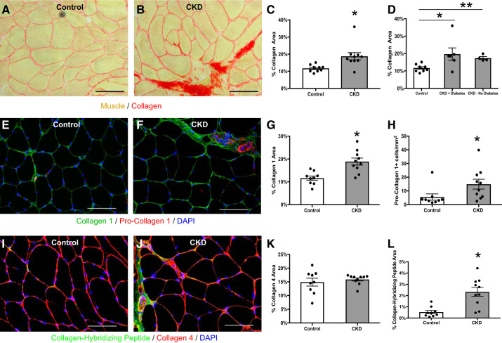
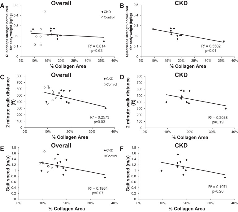
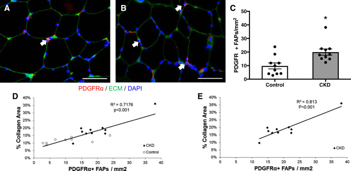
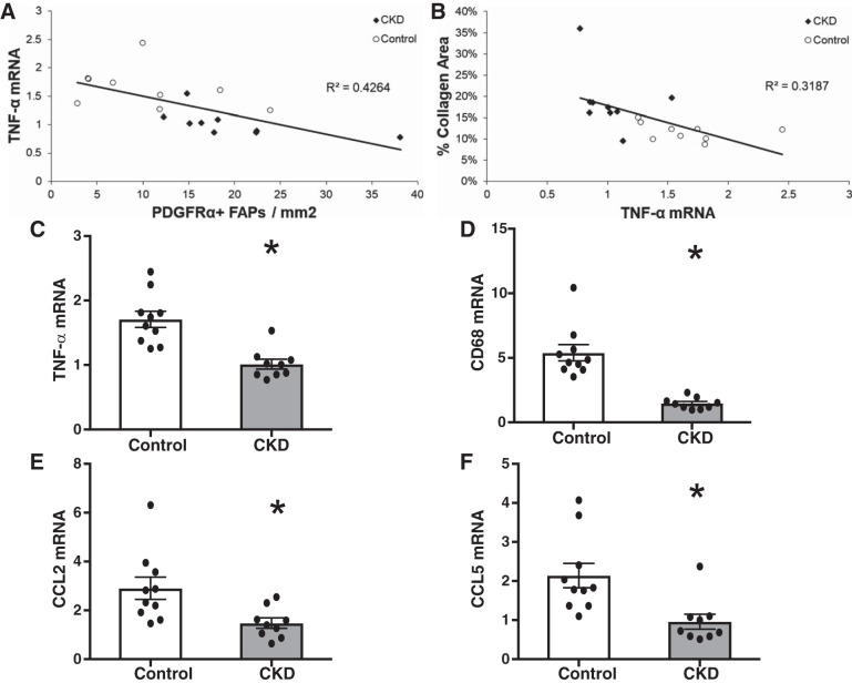
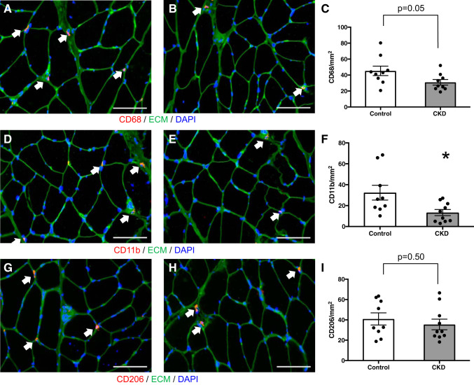
Muscle dysfunction is an important cause of morbidity among patients with chronic kidney disease (CKD). Although muscle fibrosis is present in a CKD rodent model, its existence in humans and its impact on physical function are currently unknown. We examined isometric leg extension strength and measures of skeletal muscle fibrosis and inflammation in vastus lateralis muscle from CKD patients ( n = 10) and healthy, sedentary controls ( n = 10). Histochemistry and immunohistochemistry were used to assess muscle collagen and macrophage and fibro/adipogenic progenitor (FAP) cell populations, and RT-qPCR was used to assess muscle-specific inflammatory marker expression. Muscle collagen content was significantly greater in CKD compared with control (18.8 ± 2.1 vs. 11.7 ± 0.7% collagen area, P = 0.008), as was staining for collagen I, pro-collagen I, and a novel collagen-hybridizing peptide that binds remodeling collagen. Muscle collagen was inversely associated with leg extension strength in CKD ( r = -0.74, P = 0.01). FAP abundance was increased in CKD, was highly correlated with muscle collagen ( r = 0.84, P < 0.001), and was inversely associated with TNF-α expression ( r = -0.65, P = 0.003). TNF-α, CD68, CCL2, and CCL5 mRNA were significantly lower in CKD than control, despite higher serum TNF-α and IL-6. Immunohistochemistry confirmed fewer CD68+ and CD11b+ macrophages in CKD muscle. In conclusion, skeletal muscle collagen content is increased in humans with CKD and is associated with functional parameters. Muscle fibrosis correlated with increased FAP abundance, which may be due to insufficient macrophage-mediated TNF-α secretion. These data provide a foundation for future research elucidating the mechanisms responsible for this newly identified human muscle pathology.

Keywords: chronic kidney disease; fibrosis; inflammation; skeletal muscle.


Conflict of interest statement

No conflicts of interest, financial or otherwise, are declared by the authors.

Figures

Fig. 1.
Fig. 1.
Increased collagen content within the m. vastus lateralis muscle of chronic kidney disease (CKD) patients. A and B: representative histochemical image of picro-sirius red collagen stain in control (A; n = 9) and CKD (B; n = 10) muscle biopsies. Scale bar = 100 μm. C: quantification of collagen content within the muscle represented as mean percentage of total muscle area ± SE. D: quantification of collagen content stratified by diabetes status (n = 6, CKD with diabetes; n = 4, CKD without diabetes). P values were calculated for comparisons between each CKD subgroup and control. E and F: representative immunohistochemical images demonstrating staining for collagen 1 (green), pro-collagen 1 (red), and DAPI (blue) in control (E) and CKD (F) muscle biopsies. Scale bar = 100 μm. G and H: quantification of collagen 1 content (G) and pro-collagen 1+ cells (H) within the muscle represented as mean percentage of total muscle area ± SE. I and J: representative immunohistochemical images demonstrating staining for collagen hybridizing peptide (CHP; green), collagen 4 (red), and DAPI (blue) in control (I) and CKD (J) muscle biopsies. Scale bar = 100 μm. K and L: quantification of collagen 4 content (K), and CHP binding (L) represented as mean percentage of total muscle area ± SE. *P < 0.05. **P < 0.001.
Fig. 2.
Fig. 2.
Correlation of collagen content within the m. vastus lateralis muscle with physical function in all participants (A, C, and E) and CKD patients (B, D, and F). A and B: unilateral knee extensor strength was measured in the leg to be biopsied using isometric dynamometry with a handheld dynamometer. C and D: gait speed was measured by having participants walk a 4-m course at their usual pace. E and F: endurance capacity was measured by asking participants to walk back and forth over a 50-ft course as far as possible over 2 min.
Fig. 3.
Fig. 3.
Increased fibrogenic/adipogenic progenitor (FAP) cell abundance within the m. vastus lateralis muscle of CKD patients. A and B: representative immunohistochemical image demonstrating PDGFRα+ muscle FAPs (PDGFRα+ cell surface expression surrounding DAPI+ nucleus; white arrows, red), extracellular matrix (ECM; green) and DAPI (blue) in control (A) and CKD (B) muscle biopsies. Scale bar = 50 μm. C: quantification of FAP content within the muscle represented as mean number of PDGFRα+ FAPs per total muscle area ± SE. D and E: correlation of collagen content within the m. vastus lateralis muscle with FAP content in all participants (D) and CKD patients (E). *P < 0.05.
Fig. 4.
Fig. 4.
Reduced mRNA expression of inflammatory and macrophage genes within the m. vastus lateralis muscle of CKD patients. A and B: correlation of TNF-α expression with PDGFRα+ muscle fibrogenic/adipogenic progenitor (FAP) cell abundance (A) and muscle collagen content (B). CF: mRNA expression of TNF-α, CD68, CCL2, and CCL5 in control (n = 10) and CKD (n = 9) muscle biopsies. Gene expression was normalized to RPL7 expression. Data are means ± SE. *P < 0.05.
Fig. 5.
Fig. 5.
Macrophage marker immunofluorescence within the m. vastus lateralis muscle of CKD patients and controls. A and B: representative immunohistochemical image demonstrating CD68+ muscle macrophages (white arrows, red), extracellular matrix (ECM; green), and DAPI (blue) in control (A) and CKD (B) muscle biopsies. Scale bar = 100 μm. C: quantification of CD68+ macrophage content within the muscle represented as mean number of CD68+ macrophages per total muscle area ± SE. D and E: representative immunohistochemical image demonstrating CD11b+ muscle macrophages (white arrows, red), ECM (green), and DAPI (blue) in control (D) and CKD (E) muscle biopsies. Scale bar = 100 μm. F: quantification of CD11b+ macrophage content within the muscle represented as mean number of CD11b+ macrophages per total muscle area ± SE. G and H: representative immunohistochemical image demonstrating CD206+ muscle macrophages (white arrows, red), ECM (green), and DAPI (blue) in control (G) and CKD (H) muscle biopsies. Scale bar = 100 μm. I: quantification of CD206+ macrophage content within the muscle represented as mean number of CD206+ macrophages per total muscle area ± SE. *P < 0.05.

References

    1. Ahonen RE. Striated muscle ultrastructure in uremic patients and in renal transplant recipients. Acta Neuropathol 50: 163–166, 1980. doi:10.1007/BF00692869. - DOI - PubMed
    1. Alexakis C, Partridge T, Bou-Gharios G. Implication of the satellite cell in dystrophic muscle fibrosis: a self-perpetuating mechanism of collagen overproduction. Am J Physiol Cell Physiol 293: C661–C669, 2007. doi:10.1152/ajpcell.00061.2007. - DOI - PubMed
    1. Anand S, Johansen KL, Kurella Tamura M. Aging and chronic kidney disease: the impact on physical function and cognition. J Gerontol A Biol Sci Med Sci 69: 315–322, 2014. doi:10.1093/gerona/glt109. - DOI - PMC - PubMed
    1. Bao Y, Dalrymple L, Chertow GM, Kaysen GA, Johansen KL. Frailty, dialysis initiation, and mortality in end-stage renal disease. Arch Intern Med 172: 1071–1077, 2012. doi:10.1001/archinternmed.2012.3020. - DOI - PMC - PubMed
    1. Bohannon RW, Bubela D, Magasi S, McCreath H, Wang YC, Reuben D, Rymer WZ, Gershon R. Comparison of walking performance over the first 2 minutes and the full 6 minutes of the Six-Minute Walk Test. BMC Res Notes 7: 269, 2014. doi:10.1186/1756-0500-7-269. - DOI - PMC - PubMed

Publication types

MeSH terms