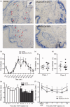
Mouse mast cells and mast cell proteases do not play a significant role in acute tissue injury pain induced by formalin
- PMID: 30280636
- PMCID: PMC6247485
- DOI: 10.1177/1744806918808161
Mouse mast cells and mast cell proteases do not play a significant role in acute tissue injury pain induced by formalin
Abstract
Subcutaneous formalin injections are used as a model for tissue injury-induced pain where formalin induces pain and inflammation indirectly by crosslinking proteins and directly through activation of the transient receptor potential A1 receptor on primary afferents. Activation of primary afferents leads to both central and peripheral release of neurotransmitters. Mast cells are found in close proximity to peripheral sensory nerve endings and express receptors for neurotransmitters released by the primary afferents, contributing to the neuro/immune interface. Mast cell proteases are found in large quantities within mast cell granules and are released continuously in small amounts and upon mast cell activation. They have a wide repertoire of proposed substrates, including Substance P and calcitonin gene-related peptide, but knowledge of their in vivo function is limited. We evaluated the role of mouse mast cell proteases (mMCPs) in tissue injury pain responses induced by formalin, using transgenic mice lacking either mMCP4, mMCP6, or carboxypeptidase A3 (CPA3), or mast cells in their entirety. Further, we investigated the role of mast cells in heat hypersensitivity following a nerve growth factor injection. No statistical difference was observed between the respective mast cell protease knockout lines and wild-type controls in the formalin test. Mast cell deficiency did not have an effect on formalin-induced nociceptive responses nor nerve growth factor-induced heat hypersensitivity. Our data thus show that mMCP4, mMCP6, and CPA3 as well as mast cells as a whole, do not play a significant role in the pain responses associated with acute tissue injury and inflammation in the formalin test. Our data also indicate that mast cells are not essential to heat hypersensitivity induced by nerve growth factor.
Keywords: Pain formalin; mast cell; protease; transgenic.
Figures


Similar articles
-
Mouse connective tissue mast cell proteases tryptase and carboxypeptidase A3 play protective roles in itch induced by endothelin-1.J Neuroinflammation. 2020 Apr 22;17(1):123. doi: 10.1186/s12974-020-01795-4. J Neuroinflammation. 2020. PMID: 32321525 Free PMC article.
-
Programming of formalin-induced nociception by neonatal LPS exposure: Maintenance by peripheral and central neuroimmune activity.Brain Behav Immun. 2015 Feb;44:235-46. doi: 10.1016/j.bbi.2014.10.014. Epub 2014 Oct 31. Brain Behav Immun. 2015. PMID: 25449583
-
The nociceptive mechanism of 5-hydroxytryptamine released into the peripheral tissue in acute inflammatory pain in rats.Eur J Pain. 2009 May;13(5):441-7. doi: 10.1016/j.ejpain.2008.06.007. Epub 2008 Jul 24. Eur J Pain. 2009. PMID: 18656400
-
Mast cell proteases as pharmacological targets.Eur J Pharmacol. 2016 May 5;778:44-55. doi: 10.1016/j.ejphar.2015.04.045. Epub 2015 May 7. Eur J Pharmacol. 2016. PMID: 25958181 Free PMC article. Review.
-
Carboxypeptidase A3-A Key Component of the Protease Phenotype of Mast Cells.Cells. 2022 Feb 6;11(3):570. doi: 10.3390/cells11030570. Cells. 2022. PMID: 35159379 Free PMC article. Review.
Cited by
-
Involvement of Mast Cells in the Pathophysiology of Pain.Front Cell Neurosci. 2021 Jun 10;15:665066. doi: 10.3389/fncel.2021.665066. eCollection 2021. Front Cell Neurosci. 2021. PMID: 34177465 Free PMC article.
-
Neurotransmitter and neuropeptide regulation of mast cell function: a systematic review.J Neuroinflammation. 2020 Nov 25;17(1):356. doi: 10.1186/s12974-020-02029-3. J Neuroinflammation. 2020. PMID: 33239034 Free PMC article.
-
The Interactions of Magnesium Sulfate and Cromoglycate in a Rat Model of Orofacial Pain; The Role of Magnesium on Mast Cell Degranulation in Neuroinflammation.Int J Mol Sci. 2023 Mar 26;24(7):6241. doi: 10.3390/ijms24076241. Int J Mol Sci. 2023. PMID: 37047214 Free PMC article.
-
Mast cell stabilizer ketotifen fumarate reverses inflammatory but not neuropathic-induced mechanical pain in mice.Pain Rep. 2021 Jun 3;6(2):e902. doi: 10.1097/PR9.0000000000000902. eCollection 2021 Jul-Aug. Pain Rep. 2021. PMID: 34104835 Free PMC article.
-
Multi-omics profiling identifies C1QA/B+ macrophages with multiple immune checkpoints associated with esophageal squamous cell carcinoma (ESCC) liver metastasis.Ann Transl Med. 2022 Nov;10(22):1249. doi: 10.21037/atm-22-5351. Ann Transl Med. 2022. PMID: 36544679 Free PMC article.
References
-
- Cox ML, Schray CL, Luster CN, Stewart ZS, Korytko PJ, M Khan KN, Paulauskis JD, Dunstan RW. Assessment of fixatives, fixation, and tissue processing on morphology and RNA integrity. Exp Mol Pathol 2006; 80: 183–191. - PubMed
-
- Story GM, Peier AM, Reeve AJ, Eid SR, Mosbacher J, Hricik TR, Earley TJ, Hergarden AC, Andersson DA, Hwang SW, McIntyre P, Jegla T, Bevan S, Patapoutian A. ANKTM1, a TRP-like channel expressed in nociceptive neurons, is activated by cold temperatures. Cell 2003; 112: 819–829. - PubMed
-
- Siebenhaar F, Magerl M, Peters EMJ, Hendrix S, Metz M, Maurer M. Mast cell–driven skin inflammation is impaired in the absence of sensory nerves. J Allergy Clin Immunol 2008; 121: 955–961. - PubMed
Publication types
MeSH terms
Substances
LinkOut - more resources
Full Text Sources
Miscellaneous

