Review
. 2019 Jan 1;99(1):21-78.
doi: 10.1152/physrev.00050.2017.

Blood-Brain Barrier: From Physiology to Disease and Back

Affiliations
Review

Blood-Brain Barrier: From Physiology to Disease and Back

Melanie D Sweeney et al. Physiol Rev. .

Abstract

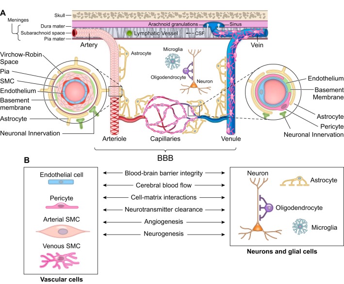
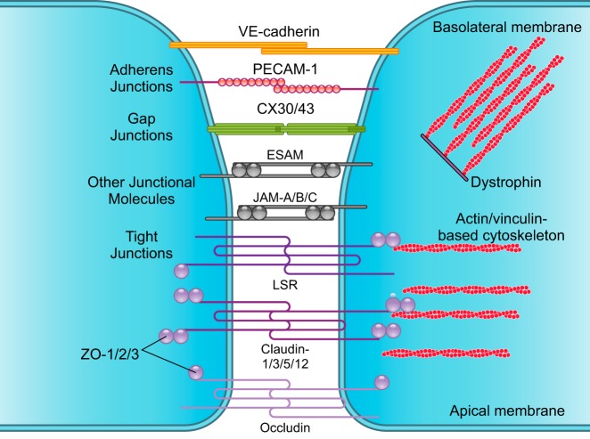
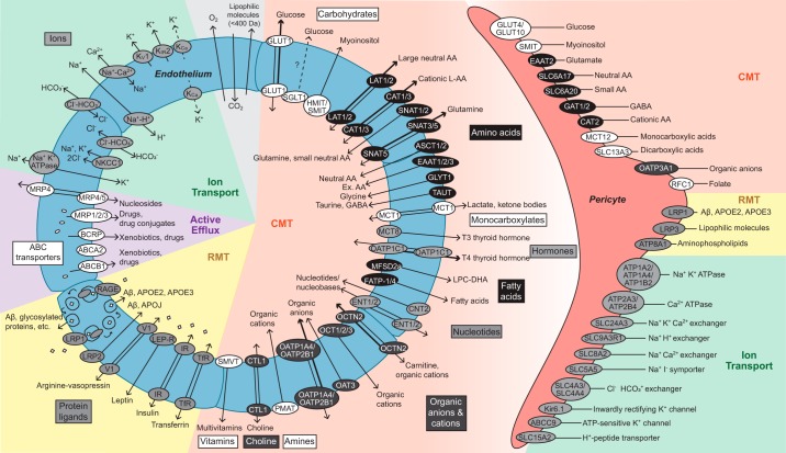
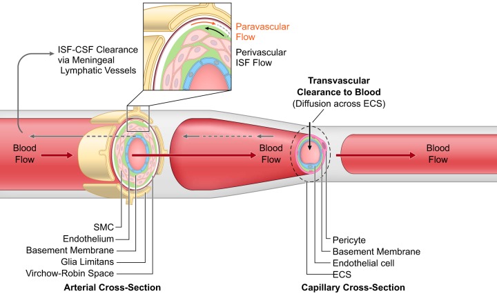
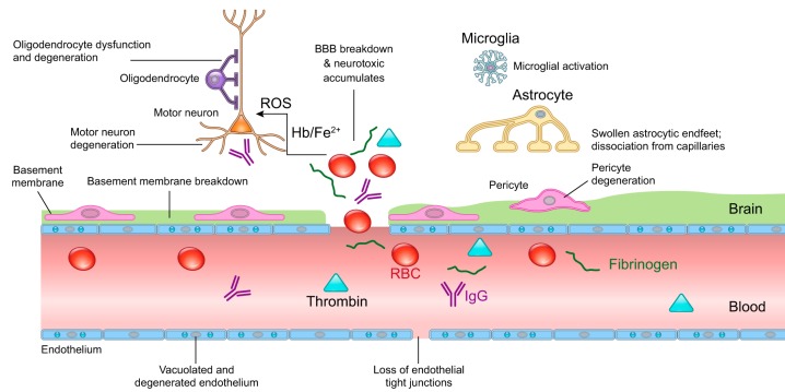
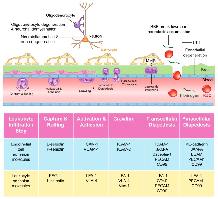
The blood-brain barrier (BBB) prevents neurotoxic plasma components, blood cells, and pathogens from entering the brain. At the same time, the BBB regulates transport of molecules into and out of the central nervous system (CNS), which maintains tightly controlled chemical composition of the neuronal milieu that is required for proper neuronal functioning. In this review, we first examine molecular and cellular mechanisms underlying the establishment of the BBB. Then, we focus on BBB transport physiology, endothelial and pericyte transporters, and perivascular and paravascular transport. Next, we discuss rare human monogenic neurological disorders with the primary genetic defect in BBB-associated cells demonstrating the link between BBB breakdown and neurodegeneration. Then, we review the effects of genes underlying inheritance and/or increased susceptibility for Alzheimer's disease (AD), Parkinson's disease (PD), Huntington's disease, and amyotrophic lateral sclerosis (ALS) on BBB in relation to other pathologies and neurological deficits. We next examine how BBB dysfunction relates to neurological deficits and other pathologies in the majority of sporadic AD, PD, and ALS cases, multiple sclerosis, other neurodegenerative disorders, and acute CNS disorders such as stroke, traumatic brain injury, spinal cord injury, and epilepsy. Lastly, we discuss BBB-based therapeutic opportunities. We conclude with lessons learned and future directions, with emphasis on technological advances to investigate the BBB functions in the living human brain, and at the molecular and cellular level, and address key unanswered questions.


Figures

FIGURE 1.
FIGURE 1.
The neurovascular unit. A: the neurovascular unit comprises vascular cells including endothelial cells and mural cells such as pericytes on brain capillaries, venules, and precapillary arterioles; vascular smooth muscle cells (SMC) on arterioles, small arteries, and veins; glial cells such as astrocytes, microglia, and olidogendrocytes; and neurons. Molecular expression patterns in endothelial and mural cells vary at different levels of vascular tree creating arterio-capillary-venous heterogeneity (zonation). At the level of penetrating arteries (left inset), endothelial cells form the inner layer of the vessel wall. The basement membrane separates endothelium from 1 to 3 layers of SMCs that are enveloped by pia. The Virchow-Robin space is between the pia and the glia limitans formed by astrocytic endfeet. At the arteriolar level, SMCs were reduced to a single layer, whereas the endothelial layer displays a continuity with the endothelium of penetrating arteries and capillaries. At the capillary level (right inset), pericytes and endothelial cells share a basement membrane and exhibit different types of cellular connections. Both the arteriolar and capillary vessel wall is covered by astrocytic endfeet. SMCs, pericytes, and astrocytes all have neuronal innervation. The blood-brain barrier (BBB) is centrally positioned within the neurovascular unit and is formed by a monolayer of tightly sealed endothelial cells extending along the vascular tree and expressing low paracellular and transcellular permeability at the level of brain capillaries and along arteriovenous axis. B: different cells of the neurovascular unit regulate BBB integrity, cerebral blood flow, extracellular matrix interactions, and neurotransmitter clearance and participate in angiogenesis and neurogenesis.
FIGURE 2.
FIGURE 2.
Brain endothelial connections. Several types of junctional molecules maintain the endothelial tight structural lining. Closest to the basolateral membrane, adherens junctions consist of vascular endothelial (VE)-cadherin and platelet endothelial cell adhesion molecule-1 (PECAM-1). Gap junctions including connexin-30 (CX30) and CX43 form hemichannels between endothelial cells. Other types of junctional molecules contribute to the tight lining including the endothelial cell adhesion molecule (ESAM) and junctional adhesion molecule (JAM)-A, -B, and -C. Closest to the apical membrane, tight junctions consist of lipolysis-stimulated lipoprotein (LSR)/angulin-1; claudin-1, -3, -5, and -12; and occludin, which limits paracellular diffusion of solutes and ions across endothelial monolayer. Zonula occludens (ZO)-1, -2, and -3 attach to claudins and occludin and bind to actin and vinculin-based cytoskeletal filaments. Dystrophin functions as a scaffold to recruit actin and vinculin, which maintains the endothelial cytoskeletal network.
FIGURE 3.
FIGURE 3.
Major blood-brain barrier transport systems. Endothelium: these include solute carrier-mediated transport (CMT), receptor-mediated transport (RMT), active efflux, and ion transport. CMT systems mediate transport of carbohydrates, amino acids, monocarboxylates, hormones, fatty acids, nucleotides, organic anions and cations, amines, choline, and vitamins with precise substrate specificity and directionality, as indicated. RMT systems transport proteins including transferrin, insulin, leptin, arginine vasopressin, amyloid-β (Aβ), glycosylated proteins, and apolipoproteins E (APOE) and J (APOJ). Active efflux includes ATP-binding cassette (ABC) transporters which transport xenobiotics, drugs, drug conjugates, and nucleosides from endothelium to blood, as indicated. Ion transport underlies the movement of Na+, K+, Cl, HCO3, H+, and Ca2+ into and out of the endothelium via ATPases, uniporters, exchangers, and symporters, as indicated. Pericytes: presently, details about pericyte transporters’ cellular polarity and precise direction(s) of transport remain elusive. CMT systems transport carbohydrates, amino acids, carboxylates, organic anions and cations, and folate. RMT system transports Aβ, APOE, lipophilic molecules, and aminophospholipids. Ion transport of Na+, K+, Cl, HCO3, H+, I, and Ca2+ occurs via ATPases, uniporters, exchangers, and symporters, as indicated. All BBB transporters indicated here are validated with RNA-sequencing and/or proteomic analysis in the rodent brain. See the main text for a more detailed discussion.
FIGURE 4.
FIGURE 4.
Perivascular and paravascular transport. Perivascular interstitial fluid (ISF) flows in the reverse direction of blood flow in the arterial vessel walls ultimately reaching cerebrospinal fluid (CSF)-filled subarachnoid spaces where ISF-CSF drains into the meningeal lymphatic vessels and cervical lymph nodes. Paravascular transport of solutes from subarachnoid spaces flows through Virchow-Robin spaces formed between pia membrane and glia limitans and is suggested to flow in the same direction as blood flow. At the capillary level, solutes diffuse across extracellular spaces (ECS) and undergo transvascular clearance to blood via transport systems as illustrated in FIGURE 3, and discussed in the text.
FIGURE 5.
FIGURE 5.
Human monogenic diseases of the blood-brain barrier. Endothelium: monogenic diseases affecting transporters include glucose transporter 1 (GLUT1) causing GLUT1 deficiency syndrome, major facilitator superfamily domain-containing protein 2a (MFSD2a) causing microcephaly 15, and monocarboxylate transporter-8 (MCT8) causing Allan-Herndon-Dudley syndrome. Cerebral cavernous malformations (CCM) are caused by mutations in endothelial proteins CCM1–3. Monogenic diseases affecting tight junctions include occludin that causes Pseudo-TORCH syndrome 1 and junctional adhesion molecule 3 (JAM3) that causes brain hemorrhagic destruction, subependymal calcification and congenital cataracts. Basement membrane: mutations affecting collagen type IV alpha 1 chain (COL4A1) and collagen type IV alpha 2 chain (COL4A2) lead to porencephaly, intracerebral hemorrhage, and cerebral small vessel disease. Pericytes: mutations in platelet-derived growth factor-BB (PDGF-BB), PDGF receptor-β (PDGFRB), solute carrier family 20 member 2 (SLC20A2), and xenotropic and polytropic retrovirus receptor 1 (XPR1) lead to idiopathic basal ganglia calcification. Vascular smooth muscle cells: mutations in notch homolog protein 3 (NOTCH3) cause cerebral autosomal dominant arteriopathy with subcortical infarcts and leukoencephalopathy (CADASIL), and mutations in HtrA serine peptidase-1 (HTRA1) protein cause cerebral autosomal recessive arteriopathy with subcortical infarcts and leukoencephalopathy (CARASIL).
FIGURE 6.
FIGURE 6.
Effects of genetic mutations carrying inheritance or increasing risk for neurodegenerative disorders on blood-brain barrier. Alzheimer’s disease (AD): APP: amyloid precursor protein (APP) vasculotropic mutations E692G (Flemish), E693Q (Dutch), E693K (Italian), E693G (Arctic), and D694N (Iowa) lead to prominent cerebral amyloid angiopathy (CAA) causing extensive cerebrovascular pathology and blood-brain barrier (BBB) breakdown in humans (orange red). Dotted boxes denote validation of BBB breakdown in transgenic rodents carrying the respective human vasculotropic mutations. APP NH2-terminal KM670/671NL (Swedish) mutations and A673 (Icelandic) mutations lead to a moderate CAA and BBB breakdown in humans (berry red). Dotted box denotes validation of BBB breakdown in transgenic animals expressing Swedish mutation. Cerebrovascular function in human carriers of APP COOH-terminal V715M (French), V715A (German), I716V (Florida), V717I (London), V717F (Indiana), and L723P (Australian) mutations has not been examined (blue; not studied). However, the BBB breakdown has been shown in transgenic models carrying Florida, London, and Indiana mutations (berry red). PSEN1: BBB breakdown and cerebrovascular dysfunction have been reported in humans carrying different PSEN1 mutations including T113–114 insertion, P117L, M139V, M146V, L153V, H163R, E184D, G209V, C260V, E280A, L282V, C285T, L420R, and ΔE9 deletion (orange red). Dotted boxes denote validation of BBB breakdown in transgenic animal models carrying the respective human PSEN1 mutations. PSEN2: the most common PSEN2 mutation N141I in humans is associated with BBB breakdown (orange red). APOE4: apolipoprotein E (APOE4), the major genetic risk factor for sporadic AD, leads to BBB breakdown in humans and transgenic models expressing human APOE4 gene (orange red, dotted box). Others: phosphatidylinositol binding clathrin assembly protein (PICALM) and clusterin (CLU) regulates clearance of amyloid-β peptide across the BBB (orange red), while sortilin-related receptor-1 (SORL1) expressed in brain endothelial cells regulates PDGF-BB and LRP1 signaling at the BBB (berry red), as shown in animal studies. Complement receptor 1 (CR1), triggering receptor expressed on myeloid cells-2 (TREM2), and bridging integrator 1 (BIN1) have not been studied for their cerebrovascular effects. Amyotrophic lateral sclerosis (ALS): transgenic rodents expressing human superoxide dismutase-1 (SOD1) G93A, G85R, and G37R mutations develop an early and pronounced BBB and blood-spinal cord barrier (BSCB) breakdown (dotted box), also confirmed in humans with familial ALS (orange red). Vascular pathology has not been studied in C9ORF72 mutation carriers (blue; not studied). Huntington’s disease: mutation in huntingtin protein causing the disease (i.e., HTT CAG repeat expansions) leads to BBB pathology in humans and animal models (orange red + dotted box). Parkinson’s disease (PD): leucine-rich repeat kinase-2 (LRRK2) mutation leads to familial PD and LRRK2 G2019S leads to a moderate BBB breakdown in humans (purple). Mutations in multi-drug resistance gene (MDR1) lead to BBB dysfunction in humans and animal models (orange red). The color scale: BBB breakdown is pronounced (orange red), moderate (berry red), modest (purple), or not studied (blue). See the main text for more detailed discussion.
FIGURE 7.
FIGURE 7.
Blood-brain barrier breakdown and dysfunction in sporadic Alzheimer’s disease. Dynamic contrast-enhanced magnetic resonance imaging (DCE-MRI) has shown blood-brain barrier (BBB) breakdown in the hippocampus in individuals with mild cognitive impairment (MCI) and in different gray and white matter regions in early Alzheimer’s disease (AD), before brain atrophy and dementia occur. Microbleeds reflecting loss of cerebrovascular integrity and BBB breakdown have been shown by T2*-MRI and susceptibility-weighted imaging (SWI)-MRI during MCI stage, which progresses and augments through early stages of AD. Fluorodeoxyglucose positron emission tomography (FDG-PET) has indicated diminished BBB GLUT1 transporter activity mediating glucose uptake by the brain before brain atrophy, dementia, or amyloid-β (Aβ) pathology. Similarly, diminished active efflux ABCB1 (P-gp) BBB transporter activity was shown by verapamil-PET in early AD. Early BBB breakdown and vascular dysfunction in MCI and AD has been confirmed by some studies by elevated levels of vascular biomarkers in cerebrospinal fluid (CSF) and blood before Aβ and tau pathology, and dementia. Neuropathological (NP) analysis of mild and advanced AD cases confirmed accumulation of perivascular blood-derived deposits including, to name a few, fibrin(ogen), thrombin, red blood cells (RBC)-derived iron-containing products that all are potentially toxic for the neural tissue. In addition, pericyte degeneration, endothelial degeneration, and brain infiltration with circulating macrophages and neutrophils were associated with BBB breakdown of AD cases on NP analysis. Diminished expression levels of BBB GLUT1 and ABCB1 (P-gp) transporters have been shown by post mortem NP analysis of AD cases, as well as downregulation of Aβ BBB clearance receptors LRP1 and ABCB1, suggesting impaired Aβ clearance. Furthermore, APOE4 carriers develop accelerated BBB breakdown associated with activation of proinflammatory cyclophilin A (CypA)-matrix metalloproteinase-9 (MMP-9) pathway at the BBB, which degrades endothelial tight junction and basement membrane proteins enhancing BBB damage. How changes in BBB permeability as measured by advanced neuroimaging techniques in the living human brain relate to disrupted structural and functional connectivity as measured by diffusion-tensor imaging (DTI)-MRI and functional MRI (fMRI), and amyloid-PET and tau-PET findings remains unclear at present.
FIGURE 8.
FIGURE 8.
Blood-brain barrier breakdown and dysfunction in sporadic Parkinson’s disease. Vascular dysfunction occurs throughout the basal ganglia of Parkinson’s disease (PD) patients, consisting of blood-brain barrier (BBB) breakdown and dysfunction, as shown by dynamic contrast-enhanced magnetic resonance imaging (DCE-MRI), T2*-MRI and susceptibility-weighted imaging (SWI)-MRI demonstrating microbleeds, and diminished active efflux of xenobiotics and other potential toxins, as indicated by verapamil-PET. Aberrant angiogenesis with increased number of endothelial cells, decreased tight junction (TJ) and adherens junction (AJ) proteins, and capillary basement membrane changes have been shown both in humans with PD and animal models. BBB breakdown can lead to neurotoxic accumulates of fibrin(ogen), thrombin, and plasmin(ogen), and red blood cell (RBC) extravasation, release of hemoglobin (Hb) and iron (Fe2+) causing reactive oxygen species (ROS), which all can injure dopaminergic neurons. Concurrently, localized to the substantia nigra pars compacta, Lewy bodies form from filamentous and oligomeric α-synuclein (α-syn) that accumulate within dopaminergic neurons. Recent studies suggested that α-syn can cross the BBB and contribute to α-syn pool in the brain, and is also cleared from brain across the BBB via LRP1-mediated transcytosis.
FIGURE 9.
FIGURE 9.
Blood-brain barrier breakdown and dysfunction in amyotrophic lateral sclerosis. Blood-brain barrier (BBB) breakdown with loss of tight junction proteins and pericyte and endothelial cell degeneration leads to red blood cells (RBCs) extravasation and perivascular accumulation of plasma-derived proteins such as fibrin(ogen), thrombin, and IgG that is found in the spinal cord and motor cortex both in humans with sporadic and familial forms of ALS, as well as in rodents expressing different human SOD1 mutations. The RBC extravasation leads to the release of neurotoxic hemoglobin (Hb), and free iron (Fe2+) causing generation of reactive oxygen species (ROS), which is toxic to motor neurons. Serum proteins such as fibrinogen can activate microglia enhancing non-autonomous motor neuron cell death. Astrocytic endfeet become swollen and dissociate from capillaries, and the perivascular space becomes enlarged and basement membrane breaks down. The effects of BBB breakdown on oligodendrocyte precursor cells that proliferate and mature oligodendrocytes that degenerate remain elusive at this time.
FIGURE 10.
FIGURE 10.
Blood-brain barrier breakdown and dysfunction in multiple sclerosis. Hallmark features of multiple sclerosis (MS) include vascular dysfunction, neuroinflammation, and oligodendrocyte degeneration causing neuronal demyelination and loss. An early blood-brain barrier (BBB) breakdown, neurotoxic fibrin(ogen) accumulation, reduced tight junction (TJ) protein expression, and endothelial degeneration are features of both human MS and animal MS models. Leukocytes infiltrate across the BBB in a multistep sequential process involving capture, rolling, activation, adhesion, crawling, and trans- or paracellular diapedesis. This process requires crosstalk between leukocytes and endothelial cells via precise molecular interactions, as illustrated in the diagram. See the main text for details. MMPs, matrix metalloproteinases; RBC, red blood cell.

Comment in

  • A definitive guide to the blood-brain barrier.
    Samson WK. Samson WK. Am J Physiol Regul Integr Comp Physiol. 2019 Jul 1;317(1):R14. doi: 10.1152/ajpregu.00073.2019. Epub 2019 Apr 3. Am J Physiol Regul Integr Comp Physiol. 2019. PMID: 30943048 Free PMC article. No abstract available.

References

    1. Abadier M, Haghayegh Jahromi N, Cardoso Alves L, Boscacci R, Vestweber D, Barnum S, Deutsch U, Engelhardt B, Lyck R. Cell surface levels of endothelial ICAM-1 influence the transcellular or paracellular T-cell diapedesis across the blood-brain barrier. Eur J Immunol 45: 1043–1058, 2015. doi: 10.1002/eji.201445125. - DOI - PubMed
    1. Abbott NJ, Patabendige AAK, Dolman DEM, Yusof SR, Begley DJ. Structure and function of the blood-brain barrier. Neurobiol Dis 37: 13–25, 2010. doi: 10.1016/j.nbd.2009.07.030. - DOI - PubMed
    1. Abbott NJ, Rönnbäck L, Hansson E. Astrocyte-endothelial interactions at the blood-brain barrier. Nat Rev Neurosci 7: 41–53, 2006. doi: 10.1038/nrn1824. - DOI - PubMed
    1. Absinta M, Ha S-K, Nair G, Sati P, Luciano NJ, Palisoc M, Louveau A, Zaghloul KA, Pittaluga S, Kipnis J, Reich DS. Human and nonhuman primate meninges harbor lymphatic vessels that can be visualized noninvasively by MRI. eLife 6: e29738, 2017. doi: 10.7554/eLife.29738. - DOI - PMC - PubMed
    1. Afonso PV, Ozden S, Cumont M-C, Seilhean D, Cartier L, Rezaie P, Mason S, Lambert S, Huerre M, Gessain A, Couraud P-O, Pique C, Ceccaldi P-E, Romero IA. Alteration of blood-brain barrier integrity by retroviral infection. PLoS Pathog 4: e1000205, 2008. doi: 10.1371/journal.ppat.1000205. - DOI - PMC - PubMed

Publication types

MeSH terms

Substances

LinkOut - more resources