. 2018 Dec 15;27(24):1702-1714.
doi: 10.1089/scd.2018.0010. Epub 2018 Nov 20.

Transplanted Human Pluripotent Stem Cell-Derived Mesenchymal Stem Cells Support Liver Regeneration in Gunn Rats

Affiliations

Transplanted Human Pluripotent Stem Cell-Derived Mesenchymal Stem Cells Support Liver Regeneration in Gunn Rats

Lucas-Sebastian Spitzhorn et al. Stem Cells Dev. .

Abstract

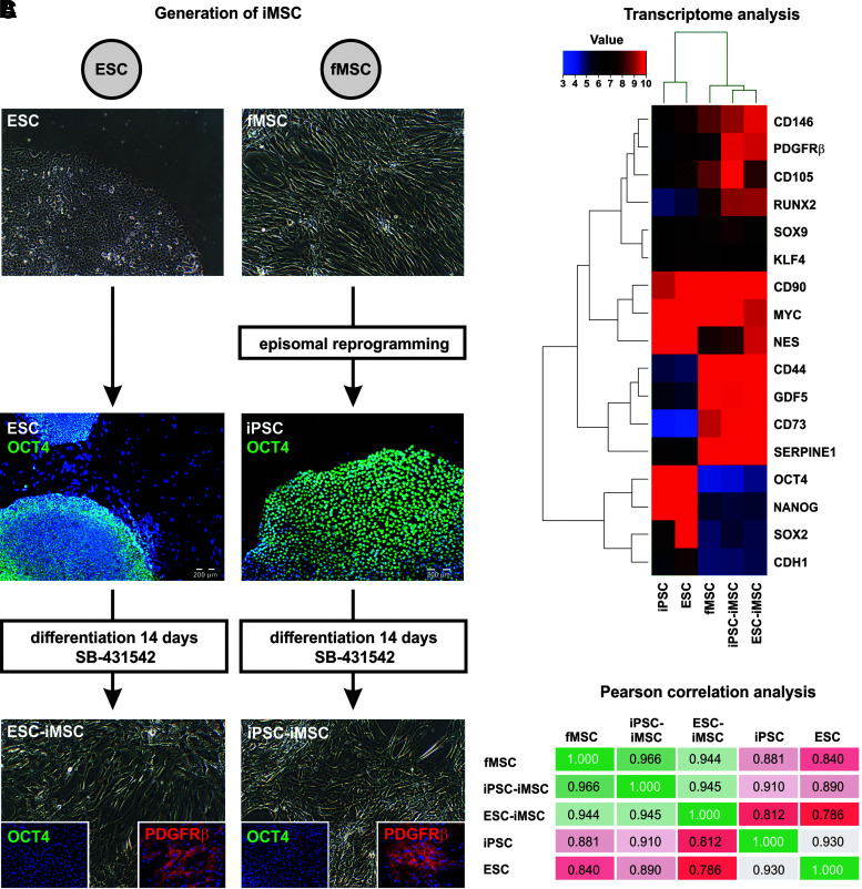
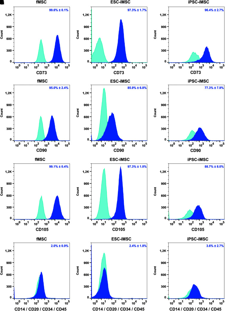
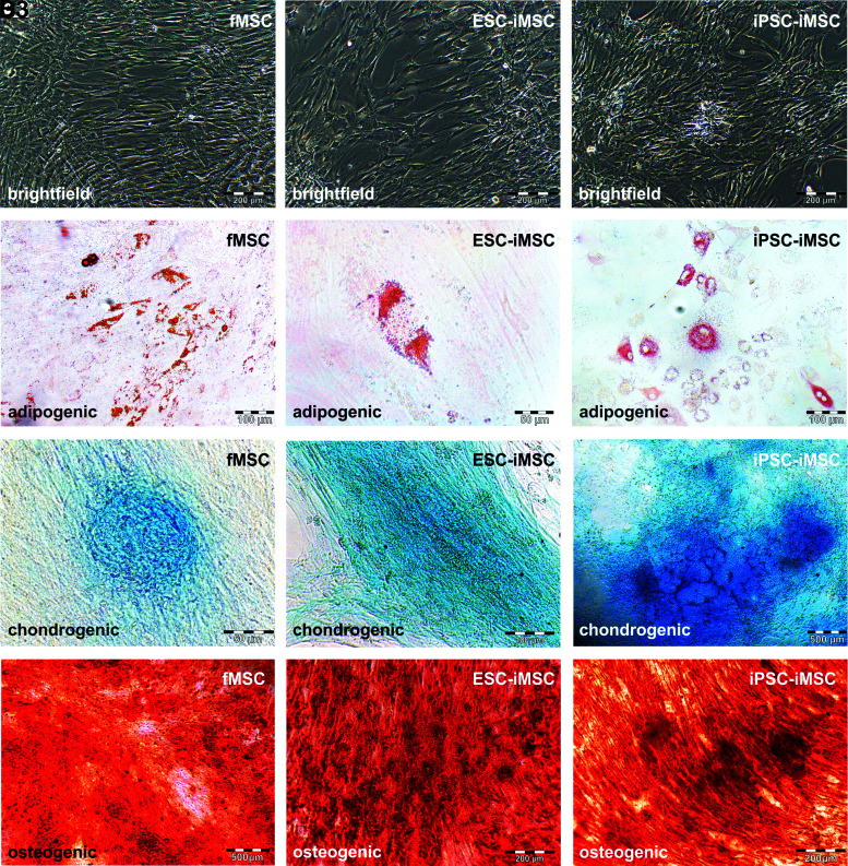
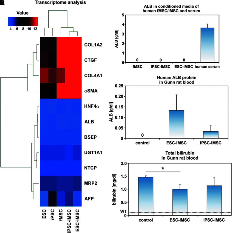
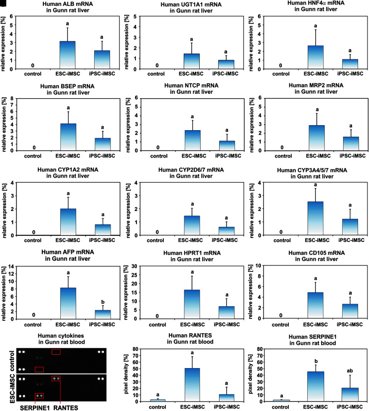
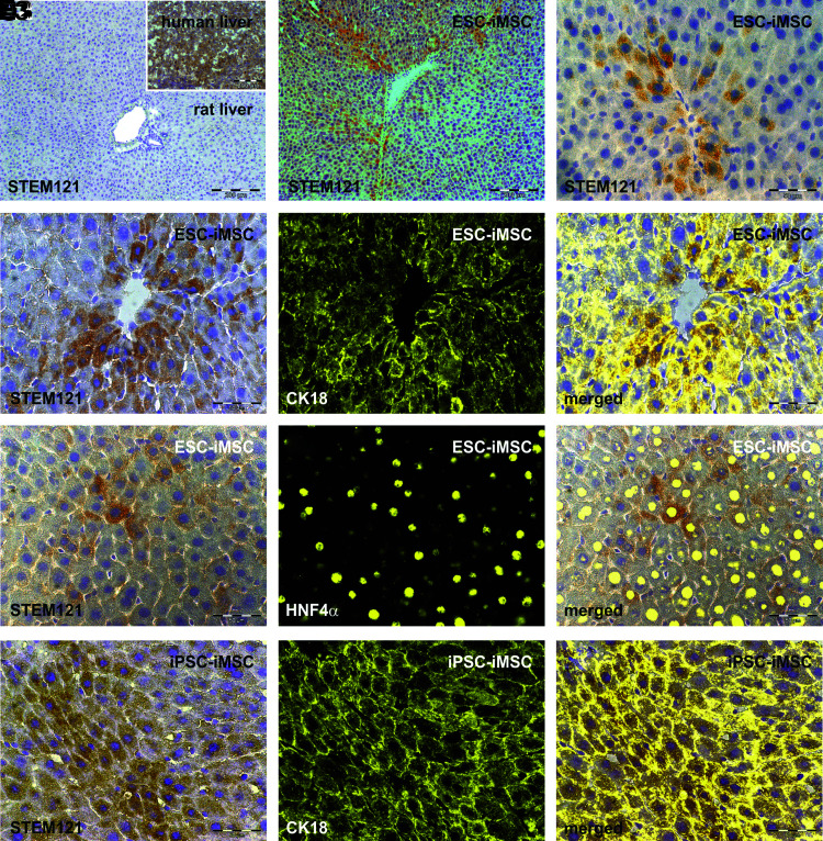
Gunn rats bear a mutation within the uridine diphosphate glucuronosyltransferase-1a1 (Ugt1a1) gene resulting in high serum bilirubin levels as seen in Crigler-Najjar syndrome. In this study, the Gunn rat was used as an animal model for heritable liver dysfunction. Induced mesenchymal stem cells (iMSCs) derived from embryonic stem cells (H1) and induced pluripotent stem cells were transplanted into Gunn rats after partial hepatectomy. The iMSCs engrafted and survived in the liver for up to 2 months. The transplanted iMSCs differentiated into functional hepatocytes as evidenced by partially suppressed hyperbilirubinemia and expression of multiple human-specific hepatocyte markers such as albumin, hepatocyte nuclear factor 4α, UGT1A1, cytokeratin 18, bile salt export pump, multidrug resistance protein 2, Na/taurocholate-cotransporting polypeptide, and α-fetoprotein. These findings imply that transplanted human iMSCs can contribute to liver regeneration in vivo and thus represent a promising tool for the treatment of inherited liver diseases.

Keywords: ESC; fetal MSC; iMSC; iPSC; liver regeneration; stem cell transplantation.


Conflict of interest statement

No competing financial interest exists.

Figures

<b>FIG. 1.</b>
FIG. 1.
Characterization of iMSCs by immunofluorescence and transcriptome analysis. (A) Scheme of iMSC generation from the human ESC line H1 and fMSCs. ESCs expressing the pluripotency-associated marker OCT4 were treated with the TGF-β receptor inhibitor SB-431542 for 14 days to facilitate their differentiation into iMSCs, concomitant with the loss of OCT4 expression and the appearance of fibroblast-shaped cells expressing the MSC marker PDGFRβ. To generate iMSCs from fMSCs, the cells were first reprogrammed into pluripotent OCT4-expressing iPSCs using plasmids encoding the pluripotency factors OCT4, SOX2, c-MYC, KLF4, NANOG, and LIN28. After treatment with SB-431542, the iPSCs differentiated into OCT4/PDGFRβ+ iMSCs as observed for ESC-iMSCs. Cell nuclei were stained with Hoechst 33258 (blue), OCT4 with FITC (green), and PDGFRβ with Cy3 (red). (B) ESC- and iPSC-derived iMSCs were characterized by gene expression array analysis (Euclidean correlation analysis). The transcriptome analysis was performed once for a representative preparation of each cell type. (C) Pearson correlation analysis of these transcriptome data revealed a high correlation (green) of both iMSCs with fMSCs, but low correlation (red) with their pluripotent precursors (ESCs and iPSCs). Scale bars indicate 200 μm. ESC, embryonic stem cell; fMSC, fetal mesenchymal stem cell; iMSC, induced mesenchymal stem cell; iPSC, induced pluripotent stem cell; OCT4, octamer-binding transcription factor 4; PDGFRβ, platelet-derived growth factor receptor β; SOX2, SRY (sex-determining region Y)-box 2; TGF-β, transforming growth factor-β.
<b>FIG. 2.</b>
FIG. 2.
Characterization of fMSCs and iMSCs by flow cytometry. The MSC markers (A–C) CD73, (D–F) CD90, and (G–I) CD105 were detectable as cell surface proteins on fMSCs and iMSCs derived from ESCs and iPSCs, whereas the expression of hematopoietic markers (J–L) CD14, CD20, CD34, and CD45 was low (dark blue). Light blue histograms represent antibody isotype controls. Representative data of repeated analysis of each cell type are shown (fMSCs n = 4; ESC-iMSCs n = 3; and iPSC-iMSCs n = 4). The percentage of positive cells for particular cell surface markers is indicated as a mean ± SEM of different analysis. SEM, standard error of mean.
<b>FIG. 3.</b>
FIG. 3.
Functional characterization of fMSCs and iMSCs. To test the developmental potential of iMSCs by qualitative assays, differentiation media for adipogenic (fMSC: n = 7; ESC-iMSC: n = 3; and iPSC-iMSC: n = 3), chondrogenic (fMSC: n = 2; ESC-iMSC: n = 3; and iPSC-iMSC: n = 2), and osteogenic (fMSC: n = 5; ESC-iMSC: n = 3; and iPSC-iMSC: n = 3) development were applied for 21 days. (A1–D1) fMSCs and iMSCs (A2–D2, ESC-iMSCs; A3–D3, iPSC-iMSCs) showed (A1–A3) fibroblast-like morphology and were able to differentiate into (B1–B3) adipocytes (Oil Red O-stained lipid droplets), (C1–C3) chondrocytes (Alcian Blue 8GX marked glycosaminoglycans in cartilage), and (D1–D3) osteoblasts (Alizarin Red stained calcium deposits). Scale bars indicate 50 μm (B2, C1), 100 μm (B1, B3, C2), 200 μm (A1–A3, D2, D3), and 500 μm (C3, D1).
<b>FIG. 4.</b>
FIG. 4.
Transplanted human iMSCs acquired hepatocyte functions in host Gunn rats. (A) Transcriptome analysis confirmed the lack of hepatocyte marker expression in pluripotent stem cells as well as fMSCs and iMSCs before transplantation, but indicated an expression of connective tissue markers and α-SMA (Euclidean correlation analysis), which are typically expressed by MSCs. The transcriptome analysis was performed once for a representative preparation of each cell type. (B) Moreover, cultured fMSCs and iMSCs did not secrete ALB into the culture medium as determined by a human-specific ALB ELISA. The normal level of ALB in human blood serum of healthy volunteers is indicated as a mean (3.6 g/dL; n = 3). (C) A differentiation of the transplanted human ESC-iMSCs and iPSC-iMSCs into hepatocytes was indicated by the presence of human ALB within the serum of Gunn rats. (D) The total bilirubin concentration in blood significantly decreased in Gunn rats after transplantation of human ESC-iMSCs, but reached no significance for iPSC-iMSCs (C, D: control n = 4; ESC-iMSC: n = 3; iPSC-iMSC: n = 3; *P < 0.05). The mean bilirubin level of normal wild-type rats (WT, 0.1 mg/dL) is indicated by a broken line [29]. ALB, albumin; α-SMA, α-smooth muscle actin; ELISA, enzyme-linked immunosorbent assay.
<b>FIG. 5.</b>
FIG. 5.
Transplanted human iMSCs successfully engrafted in Gunn rat liver and expressed hepatocyte markers. Gunn rat liver tissue and serum were analyzed 2 months after iMSC transplantation. (A) The presence of human ALB transcripts within Gunn rat liver was analyzed by qPCR. Additional hepatocyte-associated genes such as (B) UGT1A1, (C) HNF4α, (D) BSEP, (E) NTCP, (F) MRP2, (G) CYP1A2, (H) CYP2D6/7, (I) CYP3A4/5/7, and (J) AFP were found by human-specific primers for qPCR. The results were normalized to human liver samples, which were set to 100%. Thus, the bars represent proportions of the expression values of human liver. (K) Human-specific primers for HPRT1 were used to assess the total abundance of human cells derived from iMSCs. (L) The MSC marker CD105 remained detectable by human-specific primers in qPCR within the host Gunn rat livers. (M) Human RANTES and SERPINE1 were detected by protein arrays in the serum of Gunn rats transplanted with human iMSCs. A representative blot of the control and one transplantation group is shown. The three spot pairs in the corners represent protein array quality controls. Densitometric analysis of (N) RANTES and (O) SERPINE1 spots (highlighted by red boxes). The small bar of the control group represents the background pixel density (pixel density in % to the control spots). (A–I, K–L: control n = 4, ESC-iMSC, passage 10, n = 3; iPSC-iMSC, passage 10, n = 3, groups without significant differences share similar letters; P < 0.05). AFP, α-fetoprotein; BSEP, bile salt export pump; HNF4α, hepatocyte nuclear factor 4α; MRP2, multidrug resistance protein 2; NTCP, Na/taurocholate-cotransporting polypeptide; qPCR, quantitative real-time polymerase chain reaction; Ugt1A1, uridine diphosphate glucuronosyltransferase-1a1.
<b>FIG. 6.</b>
FIG. 6.
Histological analysis of human iMSC-derived hepatocytes within the Gunn rat liver. Gunn rat liver tissues were analyzed by immunohistochemistry of the human cytoplasmic protein Stem121 by DAB staining (brown) 2 months after iMSC transplantation. Liver sections of control Gunn rats 2 months after PHX showed no specific Stem121 antibody binding, whereas human liver tissue exhibited intense Stem121 DAB staining, indicating its specificity for human cells (A with insert; n = 4). Stem121 DAB staining was also combined with immunofluorescence of the hepatocyte markers CK18 or HNF4α (yellow). Human iMSC-derived hepatocytes with positive Stem121 staining were found after transplantation of (B–E3) ESC-iMSCs and (F1–F3) iPSC-iMSCs (passage 10, n = 3). The presence of Stem121 together with CK18 or HNF4α confirms the presence of hepatocytes of human origin. Scale bars indicate 50 μm (C–F3) and 200 μm (A, B). DAB, diaminobenzidine; PHX, partial hepatectomy.

Similar articles

Cited by

References

    1. Crigler JF., Jr. and Najjar VA. (1952). Congenital familial nonhemolytic jaundice with kernicterus. Pediatrics 10:169–180 - PubMed
    1. Ruud Hansen TW. (2010). Phototherapy for neonatal jaundice—therapeutic effects on more than one level? Semin Perinatol 34:231–234 - PubMed
    1. Ramy N, Ghany EA, Alsharany W, Nada A, Darwish RK, Rabie WA. and Aly H. (2016). Jaundice, phototherapy and DNA damage in full-term neonates. J Perinatol 36:132–136 - PubMed
    1. van Dijk R, Beuers U. and Bosma PJ. (2015). Gene replacement therapy for genetic hepatocellular jaundice. Clin Rev Allergy Immunol 48:243–253 - PubMed
    1. Jorns C, Nowak G, Nemeth A, Zemack H, Mörk LM, Johansson H, Gramignoli R, Watanabe M, Karadagi A, et al. (2016). De novo donor-specific HLA antibody formation in two patients with Crigler–Najjar Syndrome Type I following human hepatocyte transplantation with partial hepatectomy preconditioning. Am J Transplant 16:1021–1030 - PMC - PubMed

Publication types

MeSH terms