. 2018 Oct 3:7:e39028.
doi: 10.7554/eLife.39028.

Differing isoforms of the cobalamin binding photoreceptor AerR oppositely regulate photosystem expression

Affiliations

Differing isoforms of the cobalamin binding photoreceptor AerR oppositely regulate photosystem expression

Haruki Yamamoto et al. Elife. .

Abstract

Phototrophic microorganisms adjust photosystem synthesis in response to changes in light intensity and wavelength. A variety of different photoreceptors regulate this process. Purple photosynthetic bacteria synthesize a novel photoreceptor AerR that uses cobalamin (B12) as a blue-light absorbing chromophore to control photosystem synthesis. AerR directly interacts with the redox responding transcription factor CrtJ, affecting CrtJ's interaction with photosystem promoters. In this study, we show that AerR is translated as two isoforms that differ by 41 amino acids at the amino terminus. The ratio of these isoforms was affected by light and cell growth phase with the long variant predominating during photosynthetic exponential growth and the short variant predominating in dark conditions and/or stationary phase. Pigmentation and transcriptomic analyses show that the short AerR variant represses, while long variant activates, photosynthesis genes. The long form of AerR also activates many genes involved in cellular metabolism and motility.

Keywords: B12 photoreceptor; CrtJ/PpsR; Rhodobacter capsulatus; anoxygenic photosynthesis; genetics; genomics.


Conflict of interest statement

HY, MF, VD, CB No competing interests declared

Figures

Figure 1.
Figure 1.. AerR exists as two forms depending on the position in a cells growth cycle.
(A) R. capsulatus grown under aerobic condition and AerR-FLAG protein expression was checked at each point (1 to 14 in growth curve). (B) Western blot analysis to detect AerR-FLAG protein. Lane numbers are responding numbers in the growth curve. (C) AerR expression was checked when cells were shifted from dark to light or from light to dark after 30 and 60 min.
Figure 1—figure supplement 1.
Figure 1—figure supplement 1.. AerR also exists as two forms under photosynthetic growth conditions.
(A) R. capsulatus grown under aerobic conditions with AerR-FLAG protein expression checked at each point (1 to 13 in growth curve). (B) Western blot analysis to detect AerR-FLAG protein. Lane numbers are responding numbers in the growth curve.
Figure 1—figure supplement 2.
Figure 1—figure supplement 2.. AerR (PpaA) expression pattern in other species.
Western blot analysis of AerR(PpaA)−3xFLAG protein expression in Rhodobacter sphaeroides and Rhodospirillum centenum.
Figure 2.
Figure 2.. Mutational analysis of the internal second initiation codon, Leu41 (CTG).
All AerR mutant proteins were detected by Western blot analysis with anti-FLAG antibody. (A) Several truncated AerR proteins were expressed in R. capsulatus from internal start codon candidates, M35, M49, and M61, respectively. Frameshift mutations were introduced via one nucleotide insertions downstream of the Met1 codon. (B) One nucleotide was inserted after M35, V38, L41, T43, and V44 codons, respectively. (C) Three partial aerR sequences, that include the M1 codon to the A39, E40, or L41 codons, were fused to the bchE open reading frame that also had a FLAG epitope tag as a reporter. Translation from the M1 codon was blocked by a one nucleotide insertion after the M1 codon.
Figure 2—figure supplement 1.
Figure 2—figure supplement 1.. Summary of AerR translation start sites and AerR mutant construction.
(A) WT strain expresses both LAerR (green bar) and SAerR (red bar), from Met codon one and Leu codon 41 respectively. The two transcription start sites are indicted as a green arrow and a red arrow as defined by Sanger sequencing of the 5’-RACE experiments. Putative −35 and −10 promoter recognition sequences in blue with yellow highlighted sequences denoting FnrL binding sites. (B) An ∆SAerR strain that expresses only LAerR, contains two silent mutations indicated in red text. One is within an internal ribosome binding site used for short AerR translation and a second is at the SAerR initiation codon L41 codon. ∆LAerR strain that expresses only SAerR, has one nucleotide insertion downstream of M1 codon to stop LAerR translation. A null AerR strain was constructed that does not express any AerR isoform by insertion of one nucleotide downstream of the L45 codon. (C) AerR-FLAG expression pattern in the three AerR mutants along with a WT control.
Figure 3.
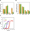
Figure 3.. Pigment levels in WT, ∆SAerR, ∆LAerR and negative control null AerR strains and their effect on photosynthetic growth.
Total pigment obtained from organic extraction from (A) aerobically or (B) photosynthetically grown WT, ∆SAerR, ∆LAerR or negative control null AerR strains that were harvested at late-exponential phase (OD = 0.6 to 0.7). Yellow bars indicate carotenoids and green bars indicate bacteriochlorophyll. (C) Photosynthetic growth of WT (blue), ∆SAerR strain (green), ∆LAerR strain (red), and the AerR null strain (purple) after shifting from dark semi-aerobic to photosynthetic illuminated conditions.
Figure 3—figure supplement 1.
Figure 3—figure supplement 1.. SAerR and LAerR phenotypes are suppressed by a CrtJ null mutation.
A crtJ gene deletion was constructed in ∆SAerR, ∆LAerR and ∆AerR mutant strains and then assayed for pigment production and photosynthetic growth. Bar graph of carotenoid (yellow bar) and bacteriochlorophyll (green bar) pigments present in dark semi-aerobically grown (A) or photosynthetically grown cells (B) harvested at late-exponential phase (OD = 0.6 to 0.7). (C) Growth curve of ∆crtJ (blue), ∆SAerR/∆crtJ (green), ∆LAerR/∆crtJ (red), Null/∆crtJ (purple), and WT control (black triangle) when shifted from dark aerobic to a light anaerobic conditions.
Figure 4.
Figure 4.. Cobalamin binding ability of SAerR protein.
(A) The insert shows a spectral scan of purified LAerR protein incubated with Ado-Cbl in dark (blue) or light (red) for 5 min followed by removal of unbound cobalamins. LAerR only binds OH-Cbl which is a product of light excitation of Ado-Cbl, thus LAerR does not bind Cbl under dark conditions. The larger spectrum is of SAerR incubated with Ado-Cbl in dark (blue) or light (red) for 5 min followed by removal of unbound cobalamins. SAerR binds both Ado-Cbl in the dark as well as light generated OH-Cbl. (B) Spectral scan of SAerR incubated with Ado-Cbl (blue), Cyano-Cbl (green), or Methyl-Cbl (pink) in dark for 5 min followed by removal of unbound cobalamins. (C) Spectral scan of purified SAerR (red), SAerR_H145A (green), and SAerR_H145A/G148E (blue) incubated with Ado-Cbl in light illumination for 5 min followed by removal of unbound cobalamins.
Figure 4—figure supplement 1.
Figure 4—figure supplement 1.. SAerR binds several kinds of cobalamin.
Spectral scan of SAerR incubated with Ado-Cbl (blue), OH-Cbl (red), Cyano-Cbl (green), or Methyl-Cbl (pink) followed by removal of unbound cobalamins. Solid lines are SAerR bound form and dotted lines are protein free of unbound cobalamins.
Figure 5.
Figure 5.. An SAerR overexpression phenotype is suppressed in vivo by mutations in the cobalamin binding motif.
(A) Spectrum of total pigment extracts from semi-aerobically grown WT strain (blue), a WT strain overexpressing SAerR (red), and a WT strain overexpressing SAerR_H145A/G148E (orange). (B) Growth curve of WT cells (blue), WT cells overexpressing SAerR (red), and WT cells overexpressing SAerR_H145A/G148E (orange). These cells were shifted from dark semi-aerobic to photosynthetic growth conditions.
Figure 6.
Figure 6.. Heat map of global expression changes in AerR and CrtJ mutant strains as measured by RNA-seq.
(A) Changes in expression occuring in the photosynthesis gene cluster region of the R. capsulatus chromosome in dark semi-aerobically grown AerR mutation strains. The ΔLAerR and AerR null strains exhibit severe reduction in photosystem transcription while the ΔSAerR exhibits an increase in expression. The reduced expression phenotypes exhibited by the ΔLAerR and AerR null strains are overcome by introduction of a crtJ deletion. (B) Similar to A with the exception that the cells were grown anaerobically under photosynthetic illuminated conditions. CrtJ binding motifs are shown as a red-dots.
Figure 7.
Figure 7.. LAerR and SAerR regulates genes in opposite direction.
Although most of the genes that are affected by disruption of LAerR do not change expression level upon disruption of SAerR, there are several genes that show an opposite change when SAerR is deleted. Examples include several carbon fixation genes, several branch chain transporters genes and TMAO reductase genes.
Figure 7—figure supplement 1.
Figure 7—figure supplement 1.. Regulation of CrtJ and AerR on the liv3 operon.
(A) ∆crtJ and ∆SAerR strains show similar liv operon expression levels (rcc03426-03433). However, ∆LaerR, aerR Null ∆aerR-∆crtJ and ∆aerR strains show highly decreased liv operon transcription under photosynthetic conditions. (B) ChIP-seq results of CrtJ and AerR in vivo binding to the liv3 operon. CrtJ and AerR co-localize to the liv promoter under both aerobic and photosynthetic conditions. Three CrtJ binding consensus sequences (TGTN12ACA) were found, with two corresponding to ChIP-seq peaks. ChIP-seq data used for this analysis was previously reported and deposited (Fang and Bauer, 2017).
Figure 8.
Figure 8.. Summary of the LAerR regulon in R. capsulatus under photosynthetic growth conditions.
LAerR activates carbon fixation, chemotaxis and motility, cobalamin biosynthesis, glycolysis and TCA cycle, heme biosynthesis, photosynthesis, ribosome and some transporters like xylose, ribulose and BCAA transporters. At the same time, LAerR represses bacterial microcompartment, TMAO reductase and C4-dicarboxylate transporter. These LAerR regulatory processes involve a direct interaction of LAerR with CrtJ.
Figure 9.
Figure 9.. Model of LAerR and SAerR function in controlling gene expression.
The results of this study, coupled with in vivo and in vitro DNA binding studies by Fang et al (Fang and Bauer, 2017), suggest that LAerR and SAerR are both capable of interacting with CrtJ. SAerR (red dot) complexed with Adenosyl- (Ado) or Methyl-cobalamin (Me:Cbl) is predominantly made under dark and/or stationary growth conditions. SAerR complexed with these biologically active cobalamin derivatives stimulates CrtJ (dark green) mediated photosystem repression. Under photosynthetic conditions, LAerR (light green dot) is the predominate form with this variant only binding hydroxyl cobalamin (OH-Cbl) that is generated via photolysis of Ado or methyl-cobalamin. LAerR bound with OH-Cbl interacts with CrtJ in a manner that promotes extensive interaction with target promoters (Fang and Bauer, 2017) activating gene expression.
Author response image 1.
Author response image 1.. Western blot analysis of WT cell and cells that express flag tagged SAerR and flag tagged SAerR containing the Cbl binding mutations.
Antibody used is to the flag epitope.

References

    1. Anders S, Pyl PT, Huber W. HTSeq--a Python framework to work with high-throughput sequencing data. Bioinformatics. 2015;31:166–169. doi: 10.1093/bioinformatics/btu638. - DOI - PMC - PubMed
    1. Armstrong GA. Genetic Analysis and Regulation of Carotenoid Biosynthesis. In: Blankenship R. E, Madigan M. T, Bauer C. E, editors. Anoxygenic Photosynthetic Bacteria. Dordrecht: Springer Netherlands; 1995. pp. 1135–1157.
    1. Babic S, Hunter CN, Rakhlin NJ, Simons RW, Phillips-Jones MK. Molecular characterisation of the pifC gene encoding translation initiation factor 3, which is required for normal photosynthetic complex formation in Rhodobacter sphaeroides NCIB 8253. European Journal of Biochemistry. 1997;249:564–575. doi: 10.1111/j.1432-1033.1997.t01-1-00564.x. - DOI - PubMed
    1. Berghoff BA, Glaeser J, Nuss AM, Zobawa M, Lottspeich F, Klug G. Anoxygenic photosynthesis and photooxidative stress: a particular challenge for Roseobacter. Environmental Microbiology. 2011;13:775–791. doi: 10.1111/j.1462-2920.2010.02381.x. - DOI - PubMed
    1. Braatsch S, Bernstein JR, Lessner F, Morgan J, Liao JC, Harwood CS, Beatty JT. Rhodopseudomonas palustris CGA009 has two functional ppsR genes, each of which encodes a repressor of photosynthesis gene expression. Biochemistry. 2006;45:14441–14451. doi: 10.1021/bi061074b. - DOI - PubMed

Publication types

MeSH terms