. 2018 Dec 1;29(25):3026-3038.
doi: 10.1091/mbc.E18-08-0514. Epub 2018 Oct 3.

Transfection of choanoflagellates illuminates their cell biology and the ancestry of animal septins

Affiliations

Transfection of choanoflagellates illuminates their cell biology and the ancestry of animal septins

David S Booth et al. Mol Biol Cell. .

Abstract

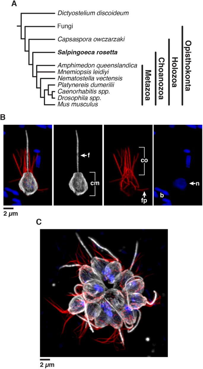
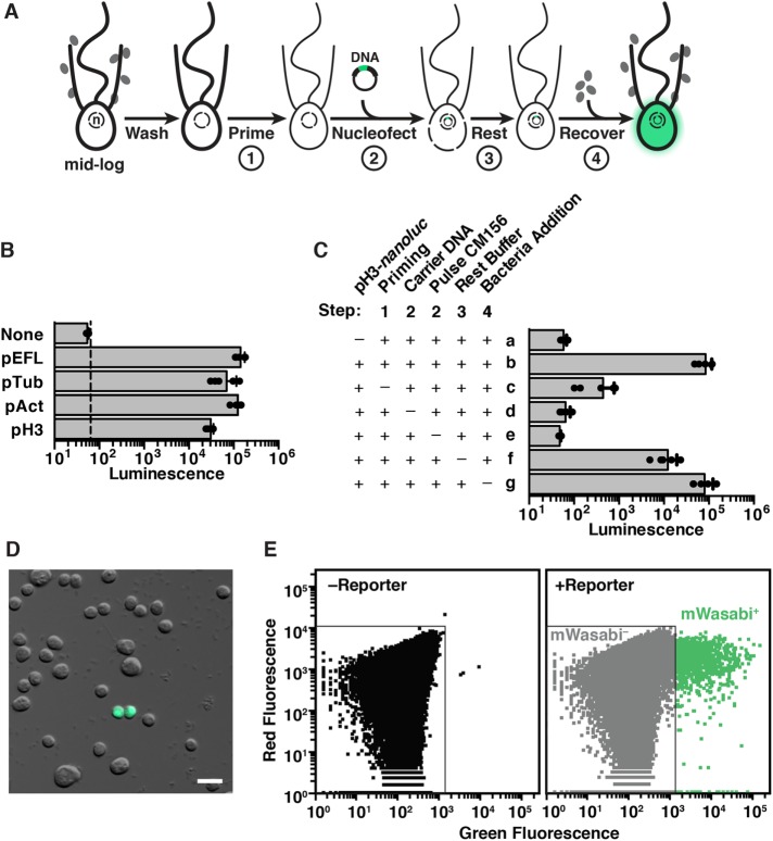
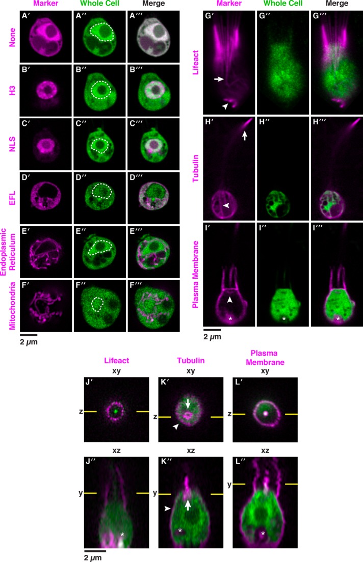
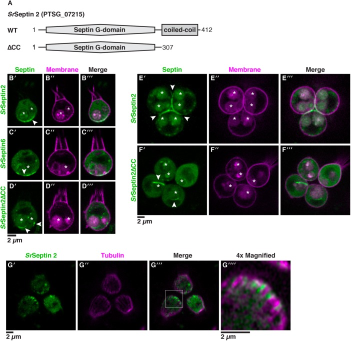
As the closest living relatives of animals, choanoflagellates offer unique insights into animal origins and core mechanisms underlying animal cell biology. However, unlike traditional model organisms, such as yeast, flies, and worms, choanoflagellates have been refractory to DNA delivery methods for expressing foreign genes. Here we report a robust method for expressing transgenes in the choanoflagellate Salpingoeca rosetta, overcoming barriers that have previously hampered DNA delivery and expression. To demonstrate how this method accelerates the study of S. rosetta cell biology, we engineered a panel of fluorescent protein markers that illuminate key features of choanoflagellate cells. We then investigated the localization of choanoflagellate septins, a family of GTP-binding cytoskeletal proteins that are hypothesized to regulate multicellular rosette development in S. rosetta. Fluorescently tagged septins localized to the basal poles of S. rosetta single cells and rosettes in a pattern resembling septin localization in animal epithelia. The establishment of transfection in S. rosetta and its application to the study of septins represent critical advances in the use of S. rosetta as an experimental model for investigating choanoflagellate cell biology, core mechanisms underlying animal cell biology, and the origin of animals.


Figures

FIGURE 1:
FIGURE 1:
Introduction to Salpingoeca rosetta, an experimentally tractable model choanoflagellate. (A) S. rosetta and other choanoflagellates are the closest living relatives of animals (Metazoa), which together with animals comprise the clade Choanozoa. (B, C) S. rosetta has a complex life history that includes single cells (B) and multicellular rosettes (C). Immuno­fluorescence in fixed, permeabilized single cells (B) highlights the diagnostic cellular architecture of choanoflagellates, including a single apical flagellum (f) made of microtubules (white) surrounded by a collar (co) filled with F-actin (red) of microvilli. Staining for tubulin also illuminates cortical microtubules (cm) that run in parallel tracks along the cell periphery from the apical to the basal poles of each cell. DNA staining (blue) highlights the choanoflagellate nucleus (n) and the nucleoids of bacterial prey (b) present in choanoflagellate cultures. In multicellular rosettes (C, stained as in B), the basal poles of cells are oriented toward the interior of the rosette and the apical flagella point outward.
FIGURE 2:
FIGURE 2:
Robust procedure for transfecting S. rosetta. (A) Summary of the stepwise procedure to transfect S. rosetta with DNA plasmids. To prepare S. rosetta for transfection, cells were harvested at mid–log phase and then washed to remove bacteria (depicted as gray ovals). S. rosetta cells (depicted with an apical collar, flagellum, and nucleus; n) were primed for nucleofection (step 1) through incubation in a buffer that degrades extracellular material. A DNA plasmid encoding a highly sensitive luciferase, nanoluc, or a fluorescent protein was then transfected into the nucleus with a nucleofector (step 2). Immediately after transfection, the cells rested in a buffer that promotes membrane closure (step 3). Finally, the cells were transferred into 1× High Nutrient Medium prepared with AK seawater for 2 d (step 4) before we assayed the expression of nanoluc or fluorescent proteins from the transfected DNA. (B) Plasmids with noncoding DNA sequences flanking the coding sequences for S. rosetta elongation factor L (pEFL), α-tubulin (pTub), β-actin (pAct), and histone H3 (pH3) genes drive the expression of a codon-optimized nanoluc reporter gene. pEFL-nanoluc, pTub-nanoluc, pAct-nanoluc, and pH3-nanoluc reporter plasmids (2.5 µg) were each transfected into S. rosetta, and the cells were subsequently assayed for luciferase expression. Each reporter produced a luminescence signal that was at least three orders of magnitude greater than the detection limit (dotted line) and significantly greater (one-way analysis of variance, p < 0.001) than the background from a negative control, in which cells were transfected with an empty pUC19 vector (None). See Materials and Methods for details on replicates and statistical tests. (C) Systematically omitting each step of the transfection procedure revealed critical steps for the delivery and expression of plasmid DNA in S. rosetta cells. As a baseline for comparison, cells with 2.5 µg of pH3-nanoluc reporter (row b) produced a luciferase signal that was three orders of magnitude greater than the background detected from cells transfected without the reporter plasmid (row a). Omitting the priming step by incubating cells in artificial seawater instead of priming buffer (row c) decreased luciferase signal by over two orders of magnitude. Nucleofection without carrier DNA (row d) or the application of the CM156 electrical pulse (row e) resulted in a complete loss of luciferase signal, indicating that both were essential for successful transfection. Directly transferring cells to sea water after nucleofection instead of to a buffer that promotes membrane resealing during the rest step (row f) decreased the luciferase signal almost 10-fold. Finally, despite the fact that most prey bacteria were washed out prior to nucleofection, addition of fresh prey bacteria did not appear to be necessary. Supplementing transfected cells with fresh prey bacteria at the start of the recovery step had seemingly little effect on transfection success (row g), probably due to the persistence of a small number of live bacteria throughout the nucleofection procedure. (D, E) Fluorescent reporters mark transfected cells. Live cells transfected with a pAct-mWasabi reporter construct could be observed by fluorescence microscopy (D) and quantified by flow cytometry (E). Untransfected cells were used to draw a gate that includes 99.99% of cells, or four SDs above the mean fluorescence value (left). The same gate was applied to a population of transfected cells (right) to categorize the mWasabi- population. Cells with higher values of green fluorescence that lay outside of the mWasabi- gate are categorized as mWasabi+. The transfection efficiency, as quantified by three independent flow cytometry experiments, was ∼1% in a population of 1 million cells.
FIGURE 3:
FIGURE 3:
Fluorescent markers illuminate the cell biology of S. rosetta in live cells. Fluorescent subcellular markers expressed from reporter plasmids in live S. rosetta cells were constructed by fusing mCherry in frame to genes encoding localization peptides and proteins (Supplemental Datasets S1 and S3). Twenty-four hours after cotransfection of cells with 5 µg of a plasmid encoding a subcellular marker fused to the mCherry protein and 5 µg of a plasmid encoding untagged mTFP1 that served as a whole cell marker, live cells were visualized by superresolution microscopy with a Zeiss LSM 880 Airyscan. The variation in localization of the whole cell mTFP1 marker stems from cell-to-cell differences in the number and localization of vacuoles, which exclude mTFP1. In panels A–I, the cells are oriented with the apical flagellum at the top and the nucleus, when included in the plane of focus (A″–F″), is indicated with a dotted white line. (A) Without localization signals (None), fluorescent proteins (mCherry, A′, and mTFP1, A″) were distributed throughout the cell with a slight enrichment in the nucleus and complete exclusion from other membrane-bound compartments. (B, C) A fusion of mCherry to the carboxy terminus of Histone H3, B′, or the amino terminus of a simian virus 40 nuclear localization signal (NLS), C′, was confined to the nucleus, whereas mCherry fused to the carboxy terminus of elongation factor L (EFL; D) was excluded from the nucleus and restricted to the cytosol. (E) The endoplasmic reticulum (ER) was highlighted by fusing the signal sequence from Rosetteless (PTSG_03555) and an ER retention sequence (HDEL from PTSG_07223) to the amino and carboxy termini of mCherry, respectively. (F) The mitochondrial network was highlighted by fusing a targeting sequence from S. cerevisiae CoxIV to the amino terminus of mCherry. (G) A Lifeact peptide fused to the amino terminus of mCherry marked filamentous actin (F-actin) that forms filipodia (arrowhead) and actin filaments in the cell body that coalesce to form the collar (arrow). (H) Fusing mCherry to the amino terminus of α-tubulin highlighted parallel tracks of microtubules (arrowhead) that extended subcortically from the apical poles to the basal poles of cells and microtubules that emerged from the apical poles of the cell bodies to form the flagella. Flagella undulate rapidly in live cells and can be difficult to image in total; in this cell the most distal tip of the flagellum is captured in the plane of focus (arrow). (I) A plasma membrane marker constructed by fusing a geranyl-geranylation sequence (PTSG_00306) to the carboxy terminus of mCherry outlined the entire cell shape, including the collar, flagellum, and cell body. The membrane marker also weakly highlighted the Golgi (arrowhead). The food vacuole (asterisk) was often visualized due to autofluorescence from ingested bacteria or through accumulation of the fluorescent markers in the food vacuole, perhaps through autophagy. (J–L) Orthogonal views along the xy and xz axes from confocal micrographs showed fine details of cell architecture that were highlighted by transfecting cells with F-actin, microtubule, and plasma membrane markers fused to mCherry (magenta). In xz views, each cell is oriented with the flagellum facing toward the top of the micrograph; flagella appeared shorter and blurred because of the sigmoidal shape of the flagellar beat. Lifeact (J) and the plasma membrane (L) markers fused to mCherry showed the microvilli (arrowheads). (K) The α-tubulin-mCherry marker showed the subcortical tracks of microtubules at the cell periphery (arrowhead) and the microtubule organizing center (arrow).
FIGURE 4:
FIGURE 4:
Septins assemble at the basal poles of S. rosetta cells. (A) SrSeptin2 has a prototypical protein domain architecture of septins, with an amino-terminal Septin G-domain that mediates filament formation and a carboxy-terminal coiled-coil domain that mediates higher-order assembly of septin filaments. To investigate the localization of SrSeptin2, we engineered fusions with mTFP1 at the amino terminus and created a truncation of the coiled-coil domain (∆CC). (B) A mTFP1-SrSeptin2 fusion protein localized to the basal pole of unicellular cells (B′, arrowhead). Cotransfecting cells with mTFP1-SrSeptin2 and a plasma membrane marker revealed SrSeptin2 distributed throughout the cytosol and enriched at the basal pole in confocal slices through the center of the cell. (C) mTFP1-SrSeptin6 mirrored the enrichment of mTFP1-SrSeptin2 at the basal pole (C′, arrowhead). The overlapping localization of SrSeptin2 and SrSeptin6 was compatible with these proteins forming heteromeric filaments with each other and other septin paralogues. (D) Consistent with the coiled-coil domain mediating the localization of septins through the formation of higher-order structures, SrSeptin2∆CC localized throughout the cytoplasm, with no visible enrichment at the basal pole. Surprisingly, the deletion also caused ectopic filaments (D′, arrowheads) to form around membrane-bound vesicles that were, based on their size and position in the cell, presumably food vacuoles. (E) In rosettes, mTFP1-SrSeptin2 localized to points of cell–cell contact corresponding to the basal poles of cells (E′; arrowhead). (F) As in single cells, mTFP1-SrSeptin2∆CC in rosettes was distributed throughout the cytosol and formed ectopic filaments (F′; arrowheads) around vacuoles. In panels E and F, S. rosetta single cells were transfected as in panels B and C, immediately induced to develop into rosettes (Woznica et al., 2016), and imaged the next day. (G) SrSeptin2 intercalated between microtubules at the basal pole of the cell. Cotransfecting cells with mTFP1-SrSeptin2 and the α-tubulin marker showed SrSeptin2 filaments intercalated between microtubules at the basal pole in confocal slices that capture the cell cortex to easily visualize microtubule tracks (G′, G″, G′″; box). G″″ shows a 4× magnification of the basal pole of a representative cell (boxed region from G′, G″, G′″). In panels B–F, autofluorescence from ingested bacteria or through accumulation of the fluorescent markers highlights the food vacuole (asterisk).

Comment in

References

    1. Abedin M, King N. (2008). The premetazoan ancestry of cadherins. Science , 946–948. - PubMed
    1. Adam JC, Pringle JR, Peifer M. (2000). Evidence for functional differentiation among Drosophila septins in cytokinesis and cellularization. Mol Biol Cell , 3123–3135. - PMC - PubMed
    1. Ai HW, Henderson JN, Remington SJ, Campbell RE. (2006). Directed evolution of a monomeric, bright and photostable version of Clavularia cyan fluorescent protein: structural characterization and applications in fluorescence imaging. Biochem J , 531–540. - PMC - PubMed
    1. Ai HW, Olenych SG, Wong P, Davidson MW, Campbell RE. (2008). Hue-shifted monomeric variants of Clavularia cyan fluorescent protein: identification of the molecular determinants of color and applications in fluorescence imaging. BMC Biol , 13. - PMC - PubMed
    1. Alegado RA, Brown LW, Cao S, Dermenjian RK, Zuzow R, Fairclough SR, Clardy J, King N. (2012). A bacterial sulfonolipid triggers multicellular development in the closest living relatives of animals. Elife , e00013. - PMC - PubMed