. 2018 Oct 3;13(10):e0204901.
doi: 10.1371/journal.pone.0204901. eCollection 2018.

Telomere shortening in breast cancer cells (MCF7) under treatment with low doses of the benzylisoquinoline alkaloid chelidonine

Affiliations

Telomere shortening in breast cancer cells (MCF7) under treatment with low doses of the benzylisoquinoline alkaloid chelidonine

Sakineh Kazemi Noureini et al. PLoS One. .

Abstract

Telomeres, the specialized dynamic structures at chromosome ends, regularly shrink with every replication. Thus, they function as an internal molecular clock counting down the number of cell divisions. However, most cancer cells escape this limitation by activating telomerase, which can maintain telomere length. Previous studies showed that the benzylisoquinoline alkaloid chelidonine stimulates multiple modes of cell death and strongly down-regulates telomerase. It is still unknown if down-regulation of telomerase by chelidonine boosts substantial telomere shortening. The breast cancer cell line MCF7 was sequentially treated with very low concentrations of chelidonine over several cell passages. Telomere length and telomerase activity were measured by a monochrome multiplex quantitative PCR and a q-TRAP assay, respectively. Changes in population size and doubling time correlated well with telomerase inhibition and telomere shortening. MCF7 cell growth was arrested completely after three sequential treatments with 0.1 μM chelidonine, each ending after 48 h, while telomere length was reduced to almost 10% of the untreated control. However, treatment with 0.01 μM chelidonine did not have any apparent consequence. In addition to dose and time dependent telomerase inhibition, chelidonine changed the splicing pattern of hTERT towards non-enzyme coding isoforms of the transcript. In conclusion, telomere length and telomere stability are strongly affected by chelidonine in addition to microtubule formation.


Conflict of interest statement

The authors have declared that no competing interests exist.

Figures

Fig 1
Fig 1. Cell viability of MCF7 cells after 48 h treatment with different concentrations of chelidonine was estimated using MTT test; mean values of four independent experiments ± SEM are shown.
Fig 2
Fig 2
A) Number of population doublings and B) doubling time after long-term treatment with chelidonine (0.01; diamonds or 0.05 μM; squares) in comparison with untreated control MCF7 cells (triangles).
Fig 3
Fig 3. T/S values of cells sequentially treated with 0.05 μM (grey) and 0.1 μM (black) chelidonine 48 h in every passage were compared with telomere length of un-treated MCF7 cells (white) which was considered as 1.
Data represented are the average of two independent experiments each in duplicates ± SD values.
Fig 4
Fig 4
A) Telomerase activity as measured by q-TRAP assay and B) hTERT transcription levels using quantitative real-time RT-PCR technique in MCF-7 cells after various time of treatment with different concentrations of chelidonine. Mean value ±SEM of three logically independent experiments each containing three samples for each point was presented. (p≤0.01 in Pair-Wise Comparisons via Tukey HSD Test unless marked as + /# /* in the plot evaluating p ≤0.05. “o “represents no significance).
Fig 5
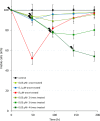
Fig 5. Cell viability of MCF7 cells after treatments (depicted with lightning bolts) 48h per passage with various concentration of chelidonine evaluated using MTT method (various concentrations and times of treatments were depicted using colour index in the chart).
The mean ± SD values of nine samples were presented.
Fig 6
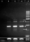
Fig 6. Alternative splice variants of hTERT from MCF-7 cells after 48 h treatment with 2 and 8 μM chelidonine in parallel with that of untreated cells.
PCR products were analysed by gel electrophoresis (3% agarose gels). The white arrows show the location of four splice variants in untreated cells. The upper band is the functional full-length hTERT (FL, 457 bp) which is followed by the three shorter non-enzyme coding variants. Lane 1: negative control, 2: treatment with 5 μM chelidonine, 3: treatment with 2 μM chelidonine, 4: untreated control and 5: 100 bp DNA marker from which 10, 10, 10, 5 and 5 μl was loaded respectively; as seen in Fig 4B the total transcription of hTERT was strongly repressed while the major isoform is minus beta. Five microliters of β2-microglobulin PCR products of the related samples have been loaded as control at bottom.

Similar articles

Cited by

References

    1. Greider CW. Telomere length regulation. Annu Rev Biochem. 1996;65: 337–65. 10.1146/annurev.bi.65.070196.002005 - DOI - PubMed
    1. Zakian VA. Structure and function of telomeres. Annu Rev Genet. 1989;23: 579–604. 10.1146/annurev.ge.23.120189.003051 - DOI - PubMed
    1. Rhodes D, Giraldo R. Telomere structure and function. Curr Opin Struct Biol. 1995; 5: 311–322. - PubMed
    1. Svenson U, Nordfjäll K. Breast cancer survival is associated with telomere length in peripheral blood cells. Cancer Res. 2008;68: 3618–3623. 10.1158/0008-5472.CAN-07-6497 - DOI - PubMed
    1. Allegra A, Innao V. Telomerase and telomere biology in hematological diseases: A new therapeutic target. Leuk Res. 2017;56: 60–74. 10.1016/j.leukres.2017.02.002 - DOI - PubMed

Publication types

MeSH terms