

Population genetic analysis of autophagy and phagocytosis genes in Drosophila melanogaster and D. simulans
- PMID: 30281656
- PMCID: PMC6169979
- DOI: 10.1371/journal.pone.0205024
Population genetic analysis of autophagy and phagocytosis genes in Drosophila melanogaster and D. simulans
Abstract
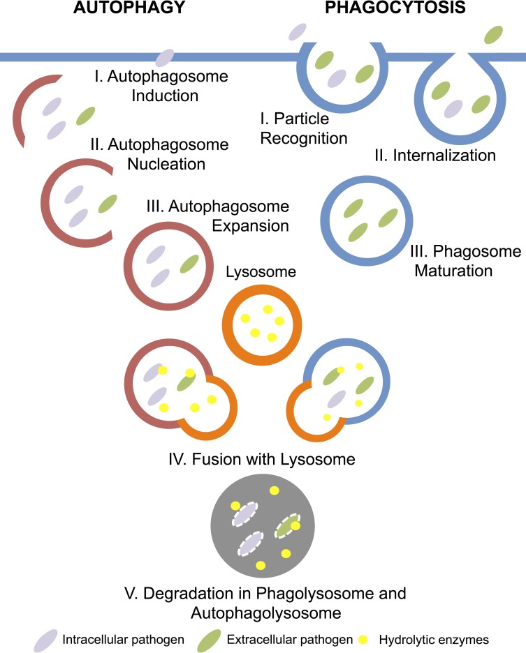
Autophagy and phagocytosis are cellular immune mechanisms for internalization and elimination of intracellular and extracellular pathogens. Some pathogens have evolved the ability to inhibit or manipulate these processes, raising the prospect of adaptive reciprocal co-evolution by the host. We performed population genetic analyses on phagocytosis and autophagy genes in Drosophila melanogaster and D. simulans to test for molecular evolutionary signatures of immune adaptation. We found that phagocytosis and autophagy genes as a whole exhibited an elevated level of haplotype homozygosity in both species. In addition, we detected signatures of recent selection, notably in the Atg14 and Ykt6 genes in D. melanogaster and a pattern of elevated sequence divergence in the genderblind (gb) gene on the D. simulans lineage. These results suggest that the evolution of the host cellular immune system as a whole may be shaped by a dynamic conflict between Drosophila and its pathogens even without pervasive evidence of strong adaptive evolution at the individual gene level.
Conflict of interest statement
The authors have declared that no competing interests exist.
Figures
Similar articles
-
A Large Panel of Drosophila simulans Reveals an Abundance of Common Variants.Genome Biol Evol. 2018 Jan 1;10(1):189-206. doi: 10.1093/gbe/evx262. Genome Biol Evol. 2018. PMID: 29228179 Free PMC article.
-
Comparative population genomics of latitudinal variation in Drosophila simulans and Drosophila melanogaster.Mol Ecol. 2016 Feb;25(3):723-40. doi: 10.1111/mec.13446. Epub 2016 Jan 18. Mol Ecol. 2016. PMID: 26523848 Free PMC article.
-
Genomics of parallel adaptation at two timescales in Drosophila.PLoS Genet. 2017 Oct 2;13(10):e1007016. doi: 10.1371/journal.pgen.1007016. eCollection 2017 Oct. PLoS Genet. 2017. PMID: 28968391 Free PMC article.
-
Patterns of polymorphism and divergence from noncoding sequences of Drosophila melanogaster and D. simulans: evidence for nonequilibrium processes.Mol Biol Evol. 2005 Jan;22(1):51-62. doi: 10.1093/molbev/msh269. Epub 2004 Sep 29. Mol Biol Evol. 2005. PMID: 15456897 Review.
-
Drosophila phagocytosis - still many unknowns under the surface.APMIS. 2011 Oct;119(10):651-62. doi: 10.1111/j.1600-0463.2011.02792.x. Epub 2011 Jul 21. APMIS. 2011. PMID: 21917002 Review.
Cited by
-
Drosophila as a Model to Study Brain Innate Immunity in Health and Disease.Int J Mol Sci. 2018 Dec 7;19(12):3922. doi: 10.3390/ijms19123922. Int J Mol Sci. 2018. PMID: 30544507 Free PMC article. Review.
-
A Population Genomic Investigation of Immune Cell Diversity and Phagocytic Capacity in a Butterfly.Genes (Basel). 2021 Feb 16;12(2):279. doi: 10.3390/genes12020279. Genes (Basel). 2021. PMID: 33669297 Free PMC article.
References
Publication types
MeSH terms
Substances
Grants and funding
LinkOut - more resources
Full Text Sources
Molecular Biology Databases
