

Structural characterization of four different naturally occurring porcine collagen membranes suitable for medical applications
- PMID: 30281664
- PMCID: PMC6169977
- DOI: 10.1371/journal.pone.0205027
Structural characterization of four different naturally occurring porcine collagen membranes suitable for medical applications
Abstract
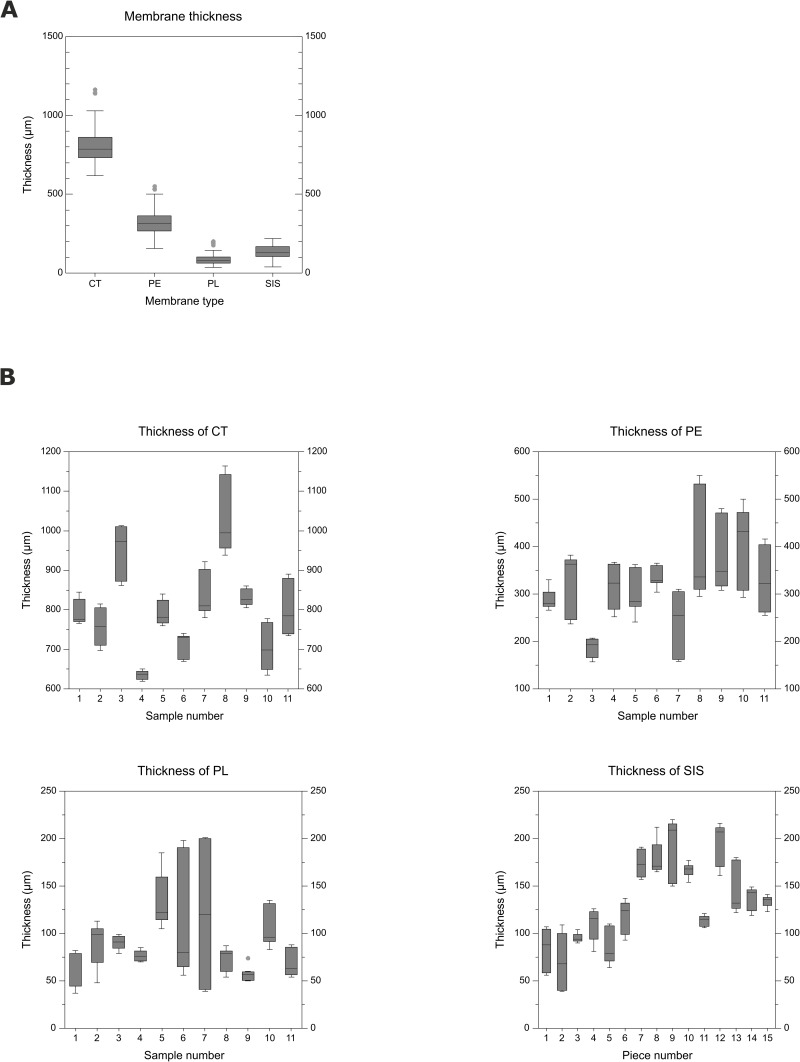
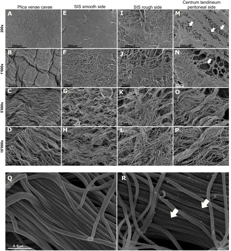
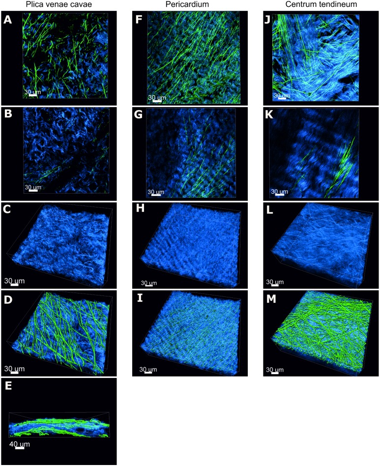
Collagen is the main structural element of connective tissues, and its favorable properties make it an ideal biomaterial for regenerative medicine. In dental medicine, collagen barrier membranes fabricated from naturally occurring tissues are used for guided bone regeneration. Since the morphological characteristics of collagen membranes play a crucial role in their mechanical properties and affect the cellular behavior at the defect site, in-depth knowledge of the structure is key. As a base for the development of novel collagen membranes, an extensive morphological analysis of four porcine membranes, including centrum tendineum, pericardium, plica venae cavae and small intestinal submucosa, was performed. Native membranes were analyzed in terms of their thickness. Second harmonic generation and two-photon excitation microscopy of the native membranes showed the 3D architecture of the collagen and elastic fibers, as well as a volumetric index of these two membrane components. The surface morphology, fiber arrangement, collagen fibril diameter and D-periodicity of decellularized membranes were investigated by scanning electron microscopy. All the membrane types showed significant differences in thickness. In general, undulating collagen fibers were arranged in stacked layers, which were parallel to the membrane surface. Multiphoton microscopy revealed a conspicuous superficial elastic fiber network, while the elastin content in deeper layers varied. The elastin/collagen volumetric index was very similar in the investigated membranes and indicated that the collagen content was clearly higher than the elastin content. The surface of both the pericardium and plica venae cavae and the cranial surface of the centrum tendineum revealed a smooth, tightly arranged and crumpled morphology. On the caudal face of the centrum tendineum, a compact collagen arrangement was interrupted by clusters of circular discontinuities. In contrast, both surfaces of the small intestinal submucosa were fibrous, fuzzy and irregular. All the membranes consisted of largely uniform fibrils displaying the characteristic D-banding. This study reveals similarities and relevant differences among the investigated porcine membranes, suggesting that each membrane represents a unique biomaterial suitable for specific applications.
Conflict of interest statement
Geistlich Pharma AG provided the salary of TM and covered the costs related to 2-photonmicroscopy. Geistlich (BS, NS) was further involved in the experimental design, data analysis, decision to publish and in the preparation of the manuscript. Internal institutional resources (government funding) covered personnel costs (JB, MHS, HM, BV, SK) as well as the expenses for laboratory consumables and scanning electron microscopy. There was no additional external funding received for this study. The authors declare no competing interests. None of the institutions involved has filed a patent application or is considering to do so. The University of Bern (Veterinary Anatomy) and Geistlich Pharma AG adopted a Research Collaboration Agreement. The commercial affiliation does not alter the authors’ adherence to all the PLOS ONE policies on sharing data and materials.
Figures
Similar articles
-
Transmural variation in elastin fiber orientation distribution in the arterial wall.J Mech Behav Biomed Mater. 2018 Jan;77:745-753. doi: 10.1016/j.jmbbm.2017.08.002. Epub 2017 Aug 5. J Mech Behav Biomed Mater. 2018. PMID: 28838859 Free PMC article.
-
Physicochemical characterization of barrier membranes for bone regeneration.J Mech Behav Biomed Mater. 2019 Sep;97:13-20. doi: 10.1016/j.jmbbm.2019.04.053. Epub 2019 May 4. J Mech Behav Biomed Mater. 2019. PMID: 31085456
-
Differences in degradation behavior of two non-cross-linked collagen barrier membranes: an in vitro and in vivo study.Clin Oral Implants Res. 2014 Dec;25(12):1403-11. doi: 10.1111/clr.12284. Clin Oral Implants Res. 2014. PMID: 25539007
-
Collagen fibers, reticular fibers and elastic fibers. A comprehensive understanding from a morphological viewpoint.Arch Histol Cytol. 2002 Jun;65(2):109-26. doi: 10.1679/aohc.65.109. Arch Histol Cytol. 2002. PMID: 12164335 Review.
-
[The three-dimensional ultrastructure of the collagen fibers, reticular fibers and elastic fibers: a review].Kaibogaku Zasshi. 1992 Jun;67(3):186-99. Kaibogaku Zasshi. 1992. PMID: 1523957 Review. Japanese.
Cited by
-
A comprehensive in vitro characterization of non-crosslinked, diverse tissue-derived collagen-based membranes intended for assisting bone regeneration.PLoS One. 2024 Jul 15;19(7):e0298280. doi: 10.1371/journal.pone.0298280. eCollection 2024. PLoS One. 2024. PMID: 39008482 Free PMC article.
-
Toughness Variations among Natural Casings: An Exploration on Their Biochemical and Histological Characteristics.Foods. 2022 Nov 26;11(23):3815. doi: 10.3390/foods11233815. Foods. 2022. PMID: 36496623 Free PMC article.
-
In Vitro Evaluation of Acellular Collagen Matrices Derived from Porcine Pericardium: Influence of the Sterilization Method on Its Biological Properties.Materials (Basel). 2021 Oct 21;14(21):6255. doi: 10.3390/ma14216255. Materials (Basel). 2021. PMID: 34771781 Free PMC article.
-
Differential Biodegradation Kinetics of Collagen Membranes for Bone Regeneration.Polymers (Basel). 2020 Jun 4;12(6):1290. doi: 10.3390/polym12061290. Polymers (Basel). 2020. PMID: 32512861 Free PMC article.
-
Synthesis and Structural Characterization of Four Different Concentrations of Ant Nest (Myrmecodia pendens) Collagen Membranes with Potential for Medical Applications.Clin Cosmet Investig Dent. 2024 May 27;16:179-189. doi: 10.2147/CCIDE.S446586. eCollection 2024. Clin Cosmet Investig Dent. 2024. PMID: 38827118 Free PMC article.
References
-
- Lee CH, Singla A, Lee Y. Biomedical applications of collagen. Int J Pharm. 2001; 221: 1–22. - PubMed
Publication types
MeSH terms
Substances
Associated data
LinkOut - more resources
Full Text Sources
Other Literature Sources
Research Materials
