

Clinical perspectives of PSMA PET/MRI for prostate cancer
- PMID: 30281701
- PMCID: PMC6142859
- DOI: 10.6061/clinics/2018/e586s
Clinical perspectives of PSMA PET/MRI for prostate cancer
Abstract
Prostate cancer imaging has become an important diagnostic modality for tumor evaluation. Prostate-specific membrane antigen (PSMA) positron emission tomography (PET) has been extensively studied, and the results are robust and promising. The advent of the PET/magnetic resonance imaging (MRI) has added morphofunctional information from the standard of reference MRI to highly accurate molecular information from PET. Different PSMA ligands have been used for this purpose including 68gallium and 18fluorine-labeled PET probes, which have particular features including spatial resolution, imaging quality and tracer biodistribution. The use of PSMA PET imaging is well established for evaluating biochemical recurrence, even at low prostate-specific antigen (PSA) levels, but has also shown interesting applications for tumor detection, primary staging, assessment of therapeutic responses and treatment planning. This review will outline the potential role of PSMA PET/MRI for the clinical assessment of PCa.
Conflict of interest statement
No potential conflict of interest was reported.
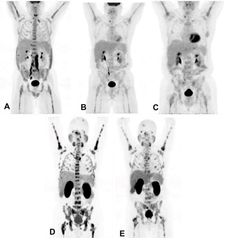
Figures


References
-
- Instituto Nacional de Cancer José Alencar Gomes da Silva (INCA) Estimativa 2016 - Incidência de Câncer no Brasil. Rio de Janeiro: INCA; 2016.
Publication types
MeSH terms
Substances
LinkOut - more resources
Full Text Sources
Medical
Research Materials
Miscellaneous
