. 2018 Oct 2;25(1):130-145.e5.
doi: 10.1016/j.celrep.2018.08.068.

Transsynaptic Binding of Orphan Receptor GPR179 to Dystroglycan-Pikachurin Complex Is Essential for the Synaptic Organization of Photoreceptors

Affiliations

Transsynaptic Binding of Orphan Receptor GPR179 to Dystroglycan-Pikachurin Complex Is Essential for the Synaptic Organization of Photoreceptors

Cesare Orlandi et al. Cell Rep. .

Abstract

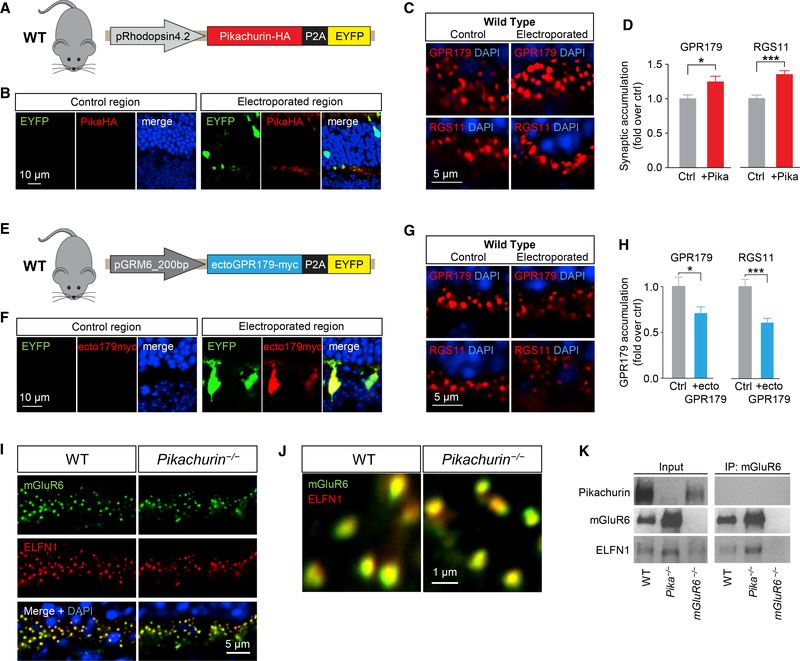
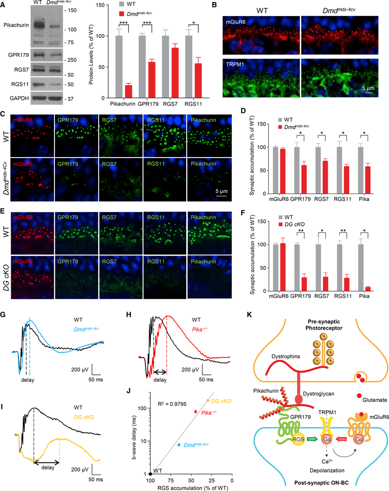
Establishing synaptic contacts between neurons is paramount for nervous system function. This process involves transsynaptic interactions between a host of cell adhesion molecules that act in cooperation with the proteins of the extracellular matrix to specify unique physiological properties of individual synaptic connections. However, understanding of the molecular mechanisms that generate functional diversity in an input-specific fashion is limited. In this study, we identify that major components of the extracellular matrix proteins present in the synaptic cleft-members of the heparan sulfate proteoglycan (HSPG) family-associate with the GPR158/179 group of orphan receptors. Using the mammalian retina as a model system, we demonstrate that the HSPG member Pikachurin, released by photoreceptors, recruits a key post-synaptic signaling complex of downstream ON-bipolar neurons in coordination with the pre-synaptic dystroglycan glycoprotein complex. We further demonstrate that this transsynaptic assembly plays an essential role in synaptic transmission of photoreceptor signals.

Keywords: HSPG; ON-bipolar cells; heparan sulfate proteoglycan; orphan receptor; retina; ribbon synapse; synaptic specificity; synaptic transmission; transsynaptic interactions; vision.


Conflict of interest statement

DECLARATION OF INTERESTS

The authors declare no competing interests.

Figures

Figure 1.
Figure 1.. Unbiased Identification of GPR158 and GPR179 as Membrane Receptors for Heparan Sulfate Proteoglycans.
(A and B) HEK293 cells were transfected with ecto-GPR158-Fc or Fc as negative control (A). The Fc proteins released in the media were isolated by incubation with protein G beads and analyzed using liquid chromatography tandem mass spectrometry (LC-MS/MS). The graph represents the interacting proteins identified specifically in the ecto-GPR-Fc pull-down experiment. DAVID analysis of the full set of identified proteins revealed the presence of 45% of secreted proteins, half of which are ECM components. The family of HSPGs is highlighted because enriched by the pull-down, and the identified members are listed in the table (B). (C and D) In vitro co-immunoprecipitation of GPR158 (C) and GPR179 (D) with representative HSPGs: GPC1, GPC5, and SDC4. HEK293 cells were transfected with the indicated myc- or HA-tagged constructs. Immunoprecipitated proteins were detected by western blotting using specific antibodies. Cells transfected with only HSPGs served as a control for non-specific binding. (E) Heparin-Sepharose pull-down from brain extract and western blot detection using a GPR158-specific antibody. Divalent cations or EDTA did not affect thepull-down. An excess of heparin (5%) was used as negative control. (F) Heparin-Sepharose pull-down from retina extract and western blot of GPR179. Divalent cations or EDTA did not affect the pull-down. An excess of heparin(5%) was used as negative control.
Figure 2.
Figure 2.. Characterization of Interactions between GPR179 and the Photoreceptor-Released HSPG Pikachurin In Vitro.
(A) Scheme of the pre-synaptic compartment at the first visual synapse. (B) In vitro co-immunoprecipitation of GPR179 with Pikachurin in HEK293 cells transfected with the indicated myc- or HA-tagged constructs. Immunoprecipitated proteins were detected by western blotting using specific antibodies. (C) Live staining of Pikachurin (red) in transfected HEK293 cells shows a predominant localization within the extracellular matrix. Incubation with conditionedmedia of HEK293 cells expressing the ecto-GPR179-Fc (green) shows co-localization with the anti-HA antibody staining (red). Negative controls are cells transfected with empty vector (pcDNA3.1) or incubated with Fc fragment. DAPI in blue. (D) Heparan sulfate modification of Pikachurin is not required for the interaction with GPR179. GPR179-myc was immunoprecipitated from cell lysate oftransfected HEK293 cells. GPR179-conjugated beads were mixed with conditioned media from cells transfected with Pikachurin-mCherry and treated with heparinase III or buffer. Cells transfected with empty vector were used as immunoprecipitation (IP) specificity control. (E) Multiple amino acid sequence alignment of pikachurin from 197 species. Identical amino acids are highlighted in orange and aligned with domain topology(below). (F) Schematics of Pikachurin structural domains and deletion constructs generated to study binding determinants to GPR179. Each construct is fused with mCherry at its C terminus. Amino acid numbers are shown at the bottom of each construct. (G) Pull-down of Pikachurin-derived mutants from HEK293 cell lysates by GPR179 ectodomain fused to an Fc fragment. Fc and mCherry are used as negativecontrols.
Figure 3.
Figure 3.. Analysis of GPR179-Pikachurin Complex Formation in the Retina.
(A) Scheme of the experimental paradigm (left). Pull-down assay of endogenous Pikachurin from retina lysates using affinity-purified ecto-GPR179-Fc expressedin transfected HEK293 cells. Fc fragment was used as negative control. (B) Confocal images of retina sections show co-localization of Pikachurin (green) and GPR179 (red) in the retina OPL. DAPI in blue. (C and D) Detection of the complex GPR179/Pikachurin (red) using proximity ligation assay (PLA) in retina cross-sections from wild-type mice. Primary antibodies were co-incubated with conditioned media of HEK293 cells expressing either the Fc fragment (C) or the ecto-GPR179-Fc as negative control (D). Dashed-line boxes indicate the region of the merged image reported with a higher magnification. PKCα (green) was used as a marker of ON-BC, and nuclei were labeled by DAPI (blue). (E) Quantification of PLA particles in the OPL or inner nuclear layer (INL) (negative control) of retina cross-sections in each condition. Data are mean ± SEM (n = 5–7, ***p < 0.001, Student’s t test). (F and G) Representative western blots (F) and quantification (G) of Pikachurin and GPR179 protein levels in retina lysates at different developmental stages. Equal amounts of total protein were loaded on a single gel, and specific antibodies were used to detect indicated proteins (n = 3). (H) Immunohistochemistry (IHC) of WT retinas showing expression pattern of Pikachurin (green) and GPR179 (red) at different developmental stages. Immunostaining of adult Gpr179nob5 retina (bottom) shows similar Pikachurin accumulation at synaptic sites. (I) Quantification of GPR179- and Pikachurin-positive puncta at the OPL during retina development in WT mice. Data are mean ± SEM (n = 3, *p < 0.05, Student’s t test).
Figure 4.
Figure 4.. Role of Pikachurin in the Targeting of the Post-synaptic Complex GPR179-RGS Proteins.
(A and B) Western blot analysis of the indicated signaling molecules in retina samples from WT and Pika−/− mice (A) and quantification normalized to GAPDH expression and reported as a percentage of WT (n = 8 WT, n = 5 Pika−/−) (B). (C) qRT-PCR of the indicated genes in WT and Pika−/− retinas (n = 5 mice/genotype). (D and E) Representative immunohistochemistry of retina sections from WT and Pika−/− mice using antibodies against mGluR6 (red), GPR179, RGS7, and RGS11 (green) (D) and related quantification (E) (n = 3). (F) In vivo retina electroporation experiments. Pika−/− mice were electroporated at P0 with the outlined photoreceptor-specific construct for Pikachurin overexpression, and the retinas were harvested after 3 weeks and prepared for IHC. (G) Representative confocal image of an individual electroporated retina immunostained with anti-HA (red) and anti-EYFP (green) antibodies. EYFP-negativeregions (left) were used as control for quantification, while EYFP-positive regions (right) represent a successful electroporation. (H and I) Representative confocal images (H) and quantification (I) of synaptic accumulation of overexpressed Pikachurin-HA in control versus electroporated regions. Five to ten different regions of retina from three different mice were used. (J and K) Representative IHC in control and electroporated retina regions in Pika−/− (J) and quantification of synaptic accumulation (K) of GPR179 (left) and RGS11 (right). DAPI in blue. Five to ten different regions of retina from three different mice were used. Data are mean ± SEM (*p < 0.05, **p < 0.01, ***p < 0.001, and ****p < 0.0001, Student’s t test).
Figure 5.
Figure 5.. Effects of Cell-Selective Pikachurin Overexpression and Dominant-Negative Blockade of GPR179-Pikachurin Complex Formation in the Retina.
(A) Schematics of the construct used for in vivo retina electroporation in WT mice. (B) IHC of WT retinas overexpressing Pikachurin-HA in photoreceptors using antibodies against HA (red) or EYFP (green). DAPI in blue. (C and D) IHC of control and electroporated regions in WT mice (C) and quantification of synaptic accumulation (D) of GPR179 (left) and RGS11 (right). DAPI in blue. Five to ten different regions of retina from three different mice were used. (E) Schematics of the dominant-negative construct expressing ectoGPR179myc under control of ON-BC-specific mGluR6 promoter used for in vivo electroporation of WT retinas. (F) IHC using antibodies against EYFP (green) and myc (red). (G and H) Representative IHC in control and electroporated retina regions in WT mice (G) and quantification of synaptic accumulation (H) of GPR179 (left) and RGS11 (right). DAPI in blue. Five to ten different regions of retina from three different mice were used. (I) IHC of mGluR6 (green) and ELFN1 (red) in retina cross-sections of WT and Pika-/−. (J) IHC of mGluR6 and ELFN1 at a higher magnification. (K) CoIP of ELFN1 and mGluR6 in retina samples from WT and Pika−/− mice. Retinas from mGluR6−/− mice were used as a control for antibody specificity. Data are mean ± SEM (*p < 0.05 and ***p < 0.001, Student’s t test).
Figure 6.
Figure 6.. Disruption of the Pre-synaptic DGC Affects Post-synaptic GPR179-RGS Complex Stability and Targeting.
(A) Representative western blots and quantification of the indicated proteins in retina samples from WT and Dmdmdx-4Cv mice. Data are mean ± SEM (n = 5 mice/genotype, *p < 0.05 and ***p < 0.001, Student’s t test). (B) Confocal images of retina sections from WT and Dmdmdx-4Cv mice stained with mGluR6 (red) and TRPM1 (green). (C) Representative confocal images of WT and Dmdmdx-4Cv mice stained with antibodies against mGluR6, GPR179, RGS7, RGS11, and Pikachurin. (D) Quantification of mGluR6, GPR179, RGS7, RGS11, and Pikachurin accumulation at the dendritic tips of ON-BCs. Nuclei were stained with DAPI (blue). Data are mean ± SEM (n = 3 mice/genotype, *p < 0.05, Student’s t test). (E) Representative IHC of retina sections from WT and DG cKO mice using antibodies against mGluR6 (red) and GPR179 or RGS7 or RGS11 (green). (F) Quantification of synaptic accumulation of the indicated proteins in WT and DG cKO mice. Data are mean ± SEM (n = 3 mice/genotype, *p < 0.05 and **p < 0.01, Student’s t test). (G–I) Representative traces of ERG responses of dark-adapted WT (black) and (G) Dmdmdx-4Cv (blue; n = 6 WT and n = 7 Dmdmdx-4Cv), (H) Pika−/− (red; n = 3 WT and n = 3 Pika−/−), and (I) DG cKO (orange; n = 3 WT and n = 3 DG cKO) mice at 1 cd s/m2. (J) Correlation between level of RGS protein accumulation (average of RGS7 and RGS11 content quantified from experiments presented in Figure 4E and in D and F) at the dendritic tips of ON-BCs and delay in b-wave implicit time (quantified and averaged from experiments presented in G–I). (K) Schematic model of the transsynaptic macromolecular complex of DGC-Pikachurin and GPR179-RGS proteins.

Similar articles

Cited by

References

    1. Ahmad R, Wojciech S, and Jockers R (2015). Hunting for the function of orphan GPCRs - beyond the search for the endogenous ligand. Br. J. Pharmacol 172, 3212–3228. - PMC - PubMed
    1. Audo I, Bujakowska K, Orhan E, Poloschek CM, Defoort-Dhellemmes S, Drumare I, Kohl S, Luu TD, Lecompte O, Zrenner E, et al. (2012). Whole-exome sequencing identifies mutations in GPR179 leading to autosomal-recessive complete congenital stationary night blindness. Am. J.Hum. Genet 90, 321–330. - PMC - PubMed
    1. Baeg GH, Lin X, Khare N, Baumgartner S, and Perrimon N (2001). Heparan sulfate proteoglycans are critical for the organization of the extracellular distribution of Wingless. Development 128, 87–94. - PubMed
    1. Bargmann CI, and Marder E (2013). From the connectome to brain function.Nat. Methods 10, 483–490. - PubMed
    1. Bishop JR, Schuksz M, and Esko JD (2007). Heparan sulphate proteoglycans fine-tune mammalian physiology. Nature 446, 1030–1037. - PubMed

Publication types