Review
. 2018 Oct 3;8(10):180135.
doi: 10.1098/rsob.180135.

Into the breach: how cells cope with wounds

Affiliations
Review

Into the breach: how cells cope with wounds

Mitsutoshi Nakamura et al. Open Biol. .

Abstract

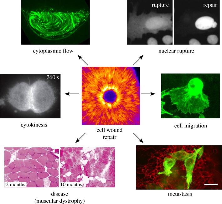
Repair of wounds to individual cells is crucial for organisms to survive daily physiological or environmental stresses, as well as pathogen assaults, which disrupt the plasma membrane. Sensing wounds, resealing membranes, closing wounds and remodelling plasma membrane/cortical cytoskeleton are four major steps that are essential to return cells to their pre-wounded states. This process relies on dynamic changes of the membrane/cytoskeleton that are indispensable for carrying out the repairs within tens of minutes. Studies from different cell wound repair models over the last two decades have revealed that the molecular mechanisms of single cell wound repair are very diverse and dependent on wound type, size, and/or species. Interestingly, different repair models have been shown to use similar proteins to achieve the same end result, albeit sometimes by distinctive mechanisms. Recent studies using cutting edge microscopy and molecular techniques are shedding new light on the molecular mechanisms during cellular wound repair. Here, we describe what is currently known about the mechanisms underlying this repair process. In addition, we discuss how the study of cellular wound repair-a powerful and inducible model-can contribute to our understanding of other fundamental biological processes such as cytokinesis, cell migration, cancer metastasis and human diseases.

Keywords: Rho GTPases; actomyosin; calcium; membrane trafficking; plasma membrane repair; single cell wound repair.


Conflict of interest statement

The authors declare no competing interests.

Figures

Figure 1.
Figure 1.
Schematic of major steps in cell wound repair. (a) Cross section of unwounded cell showing intact plasma membrane and underlying cortical cytoskeleton. (b) Cells with a membrane lesion showing influx of calcium ions (left panel) or reactive oxygen species (right panel) that oxidize cellular components. Influx of these ions each start signalling cascades that initiate cellular wound repair processes. (c) Cells quickly close wounds, either through a vesicular membrane patch (left panel), or by endocytosis/exocytosis of the area of membrane containing the lesion (right panel). (d) Actin based cytoskeleton works to close cellular wounds by an actomyosin contractile ring (left panel), actin treadmilling where the leading edge closest to the wound is the site of F-actin assembly and the trailing edge distal to the wound has F-actin disassembly (middle panel), or by a transient actin patch that accumulates under the membrane lesion (right panel). (e) Cells with repaired wounds must remodel their plasma membrane and cortical cytoskeleton to their pre-wounded state. The fate of the transient membrane patch is not well studied, but it is thought to be removed from the membrane by either endocytosis or exocytosis (left panel). In addition, components of the actin cytoskeleton, which are enriched at the wound site are thought to be either endocytosed or exocytosed in order to return that region to its pre-wounded state (right panel). (f) Cell with completely healed wound—plasma membrane and underlying cortical cytoskeleton have been returned to their unwounded state.
Figure 2.
Figure 2.
Calcium influx and oxidation are the most upstream events in cell wound repair. (a) Ca2+ dependence of cell wound repair. An FM1-43 labelled sea urchin egg was wounded twice with a laser. The first wound (arrow; upper left panel) was generated in the presence of calcium and exhibits a robust repair response (upper right panel). The sea urchin egg was moved to calcium-free sea water and a second wound (arrow; lower left panel) was then generated. In the absence of calcium no repair is initiated and cytoplasm can be seen flowing out of the wound (lower right panel). Adapted by permission from Springer Nature and Copyright Clearance Center: McNeil & Kirchhausen [4] (Copyright © 2005). (b,c) Scanning electron micrographs of sea urchin eggs in the presence (b) or absence (c) of calcium upon wounding. Wounds were generated mechanically using a needle. In the presence of calcium, vesicles (arrowhead) are recruited to the wound where they fuse to each other resulting in the formation of large vesicles. In the absence of calcium, no large vesicles are formed. Adapted by permission from Springer Nature and Copyright Clearance Center: McNeil & Baker [47] (Copyright © 2001). (d,e) Recruitment of activated Rho family GTPases in Xenopus oocytes upon wounding in the presence (d) or absence (e) of calcium. Activity biosensors for Rho (rGBD) and Cdc42 (wGBD) only exhibit distinct concentric ring patterns around the wound in the presence of calcium. Republished with permission of The Rockefeller University Press, from Benink & Bement; permission conveyed through Copyright Clearance Center, Inc. [24] (Copyright © 2005). (f,g) MG53 protein tethered to vesicles and membrane accumulates at wounds (arrowheads) even in the absence of calcium. Adapted by permission from Springer Nature and Copyright Clearance Center: Cai et al. [13] (Copyright © 2009). (h) Schematic depicting the role of oxidation in cell wound repair. Vesicles coated with MG53 protein are recruited to wounds where the MG53 proteins attach to each other through oxidation-dependent disulfide bond formation.
Figure 3.
Figure 3.
Mechanisms of plasma membrane (PM) repair. (a,b) Single cell repair in a sea urchin embryo. PM repair progression in a sea urchin egg after wound induction by a polylysine coated microneedle. (a) Sea urchin eggs were immersed in salt water containing 100 µg of fluorescein stachyose (FS). Lack of FS entry into the wounded egg indicates rapid plasma membrane resealing. (b) Sea urchin eggs establish a boundary at the wound site suggesting processive PM repair. Republished with permission of The Rockefeller University Press, from Terasaki et al.; permission conveyed through Copyright Clearance Center, Inc. [11] (Copyright © 1997). (c) Time-lapse micrographs of Xenopus oocytes wounded in the presence of dextran (green) after being stained with the PM marker, rhodamine B chloride (R18, red). Vesicle fusion and rupture along the wound edge establishes a single lipid bilayer (arrowheads at 10 s and 12 s show adjacent vesicle–vesicle fusion along the wound edge followed by rupture). (d) Wounding of Xenopus oocytes in the presence of dextran. Influx of dextran through the open central wound area (arrowheads) is observed by 6 s post-wounding. At later time points, dextran puncta are observed at the wound periphery of the initial wound opening indicative of exocytosis. (e) eGFP-human Dysferlin injected into Xenopus oocytes is recruited to a ring-like structure at the wound periphery and intracellular compartments upon wounding. Images (c–e) republished with permission of American Society of Cell Biology, from Davenport et al.; permission conveyed through Copyright Clearance Center, Inc. [20] (Copyright © 2016). (f) Dysferlin-eGFP (green) co-localizes with lysosomes (red) after mechanical wounding of L6 myotubes. Adapted from McDade & Michele, by permission of Oxford University Press [61] (Copyright © 2014). (g) Laser wounding of the Drosophila embryo shows annexin B9 (green) recruitment within the wound area. Image provided by M. Nakamura. (h) MCF cells showing annexin A6 and annexin A4 recruitment upon wounding. Arrows indicate the site of wounding (upper panels); annexin A6 showed an immediate recruitment followed by annexin A4 at 5 s. Adapted from fig. 6 in Boye et al.; Creative Commons license: https://creativecommons.org/licenses/by/4.0/ [62] (Copyright © 2017). (i) Myotubes were irradiated at the plasma membrane (arrow) in buffer containing 1 mM Ca2+ and 5 µg of FM1-43 dye. Annexin A5 deficient myotubes show an increase in FM1-43 uptake when compared to the control at 120 s. Adapted and reprinted from Carmeille et al., with permission from Elsevier [63] (Copyright © 2016).
Figure 4.
Figure 4.
Cytoskeletal responses in cell wound repair. (a,b) Confocal XY projection at 180 s post-wounding (a) and kymograph (b) of NC4-staged Drosophila embryo expressing a GFP-actin reporter. Actin accumulates in two regions adjacent to the wound: a highly-enriched actin ring abutting the wound edge (red bracket), and an elevated actin halo encircling the actin ring (yellow circle). Membrane plug is also indicated. Adapted and republished with permission of The Rockefeller University Press, from Nakamura et al.; permission conveyed through Copyright Clearance Center, Inc. [81] (Copyright © 2017). (c–e) XY projections of the surface of a laser-wounded Xenopus oocyte displaying concentric rings of RhoA activity (green) (c), Cdc42 activity (green) alongside injected fluorescent actin (red) (d), and RhoA activity (red) overlaid with Cdc42 activity (green) (e). Republished with permission of The Rockefeller University Press, from Benink & Bement; permission conveyed through Copyright Clearance Center, Inc. [24] (Copyright © 2005). (f–h) XY and YZ projections alongside localized staining intensities across the wound midline of wounded Drosophila syncytial embryos for Rac1 (green) and Rho1 (red) (f), Rho1 (green) and Cdc42 (red) (g) and Rac1 (red) and Cdc42 (green) (h). Positions of GTPase recruitment (green or red arrowheads) and of GTPase co-localization (white arrows) are indicated. Adapted and reprinted from Abreu-Blanco et al., with permission from Elsevier [27] (Copyright © 2014). (i) XY views and kymograph of laser-wounded Drosophila syncytial embryo expressing fluorescent actin reporters in the presence of Y27632. Adapted and republished with permission of The Rockefeller University Press, from Abreu-Blanco et al.; permission conveyed through Copyright Clearance Center, Inc. [26] (Copyright © 2011). (j) Time lapse images at 1 and 2 min after laser-wounded Xenopus oocytes following injection with control or Y27632 (Rok kinase inhibitor that prevents myosin-II activity) alongside a fluorescent actin reporter (left). Brightest points projection across experimental time representing cortical flow alongside a kymograph demonstrating wound closure (right). Yellow line at time 00:00 represents position of kymograph. Yellow line in kymograph identifies position of the leading edge. Adapted and reprinted from Burkel et al., with permission from Elsevier [25] (Copyright © 2012). (k) Schematic depicting XY view of actomyosin dynamics following wounding in the actin treadmilling and contraction wound closure models.
Figure 5.
Figure 5.
Beyond cellular repair. The study of single cell wound healing can inform many different fields of biology, from questions in basic science to the pathology of certain diseases. Cell Wound Repair: Single cell wound repair, shown here in a Drosophila embryo, requires the coordination of many cytoskeletal components, including spatio-temporal patterning of actin at the wound site. Image provided by M. Nakamura. Cytokinesis: Dividing cells require contraction of an actomyosin ring at the cell equator, shown here in human retinal cells undergoing cytokinesis. Adapted from fig. 4a in Spira et al.; Creative Commons license: https://creativecommons.org/licenses/by/4.0/ [114] (Copyright © 2017). Cytoplasmic flows: Cortical and cytoplasmic flows are responsible for bringing cellular components to sites where they are needed. During ooplasmic streaming in a developing Drosophila oocyte, cytoplasmic flows ensure that cellular content is evenly distributed. Image provided by J. Decker. Nuclear Rupture: Nuclei must faithfully repair after nuclear envelope rupture to preserve genomic integrity and this process may share protein components with cellular membrane repair. Nuclei from U2OS cells expressing GFP with a nuclear localization sequence undergoing nuclear rupture and repair. Adapted and republished with permission of The Rockefeller University Press, from Hatch & Hetzer; permission conveyed through Copyright Clearance Center, Inc. [115] (Copyright © 2016). Cell Migration: Cells, like this migratory haemocyte (macrophage) from a Drosophila larva, move throughout their environment by rapidly altering their cytoskeleton. Image provided by S. Parkhurst. Metastasis: Melanoma cells (green) spreading in a zebra fish larval hind-brain (red). In order for cells to invade new tissues, they must navigate a dense extracellular matrix while maintaining membrane integrity. Reprinted from Roh-Johnson et al., with permission from Elsevier [116] (Copyright © 2017). Disease: Skeletal muscle—a tissue that undergoes near constant wear and tear—from Dysferlin null mice, showing defective membrane repair in response to stress contributes to cell death. Adapted by permission from Springer Nature and Copyright Clearance Center: Bansal et al. [34] (Copyright © 2003).

Similar articles

Cited by

References

    1. McNeil PL, Steinhardt RA. 1997. Loss, restoration, and maintenance of plasma membrane integrity. J. Cell Biol. 137, 1–4. (10.1083/jcb.137.1.1) - DOI - PMC - PubMed
    1. Sonnemann K, Bement W. 2012. Wound repair: toward understanding and integration of single-cell and multicellular wound responses. Annu. Rev. Cell Dev. Biol. 27, 237–263. (10.1146/annurev-cellbio-092910-154251) - DOI - PMC - PubMed
    1. Cooper ST, McNeil PL. 2015. Membrane repair: mechanisms and pathophysiology. Physiol. Rev. 95, 1205–1240. (10.1152/physrev.00037.2014) - DOI - PMC - PubMed
    1. McNeil P, Kirchhausen T. 2005. An emergency response team for membrane repair. Nat. Rev. Mol. Cell Biol. 6, 499–505. (10.1038/nrm1665) - DOI - PubMed
    1. Moe A, Golding AE, Bement WM. 2015. Cell healing: calcium, repair and regeneration. Semin. Cell Dev. Biol. 45, 18–23. (10.1016/j.semcdb.2015.09.026) - DOI - PMC - PubMed

Publication types

LinkOut - more resources