. 2018 Nov 27;92(24):e00798-18.
doi: 10.1128/JVI.00798-18. Print 2018 Dec 15.

Extracellular Hepatitis B Virus RNAs Are Heterogeneous in Length and Circulate as Capsid-Antibody Complexes in Addition to Virions in Chronic Hepatitis B Patients

Affiliations

Extracellular Hepatitis B Virus RNAs Are Heterogeneous in Length and Circulate as Capsid-Antibody Complexes in Addition to Virions in Chronic Hepatitis B Patients

Lu Bai et al. J Virol. .

Abstract

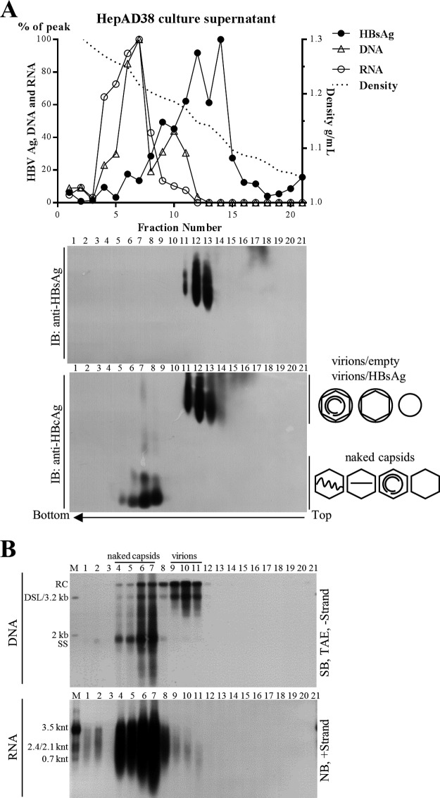
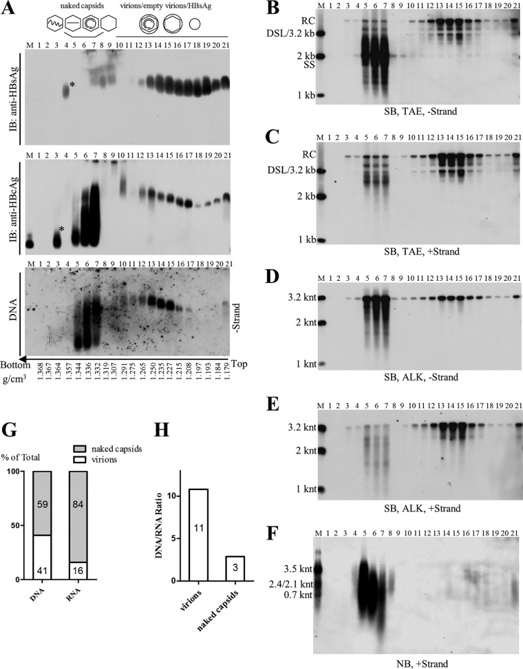
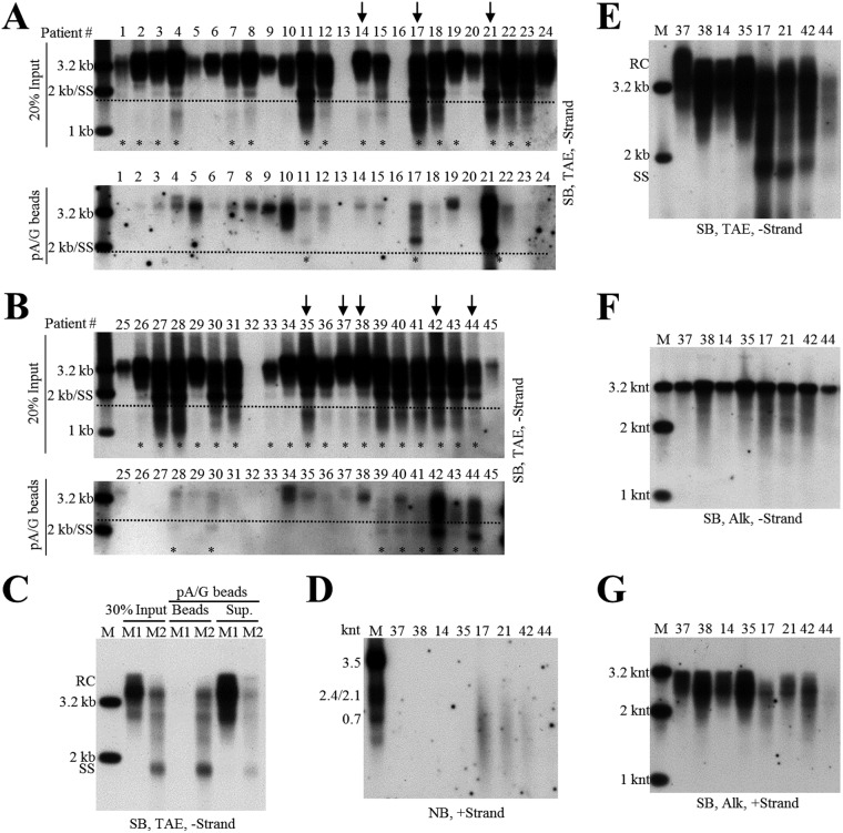
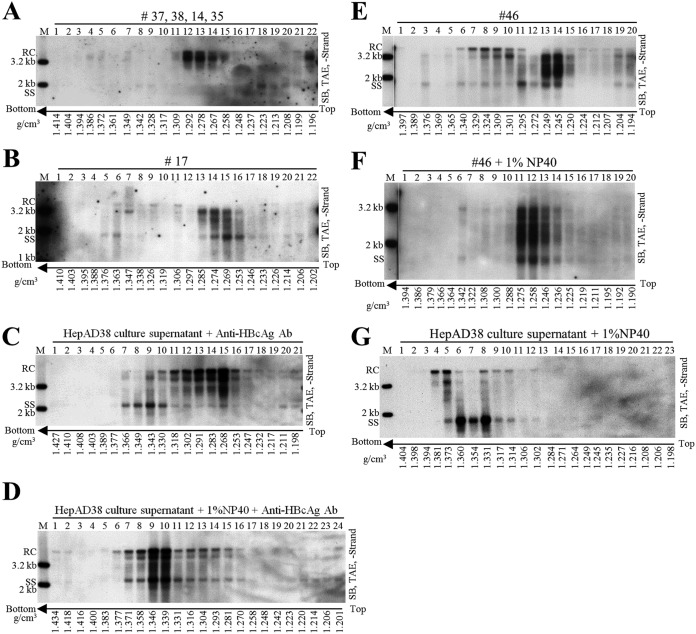
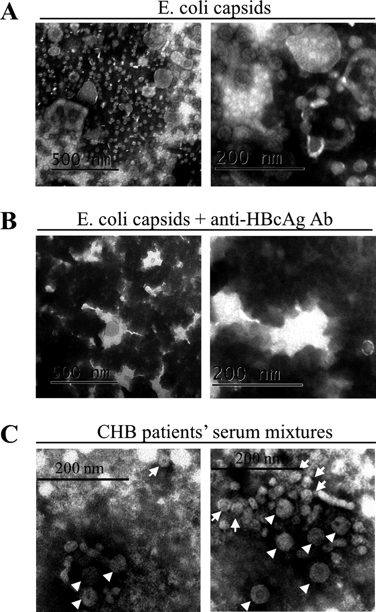
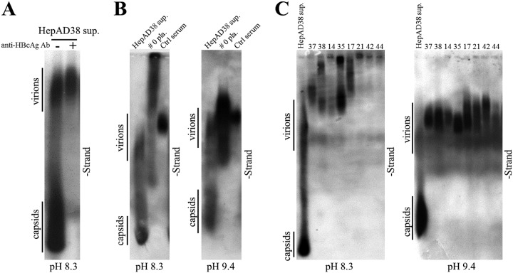
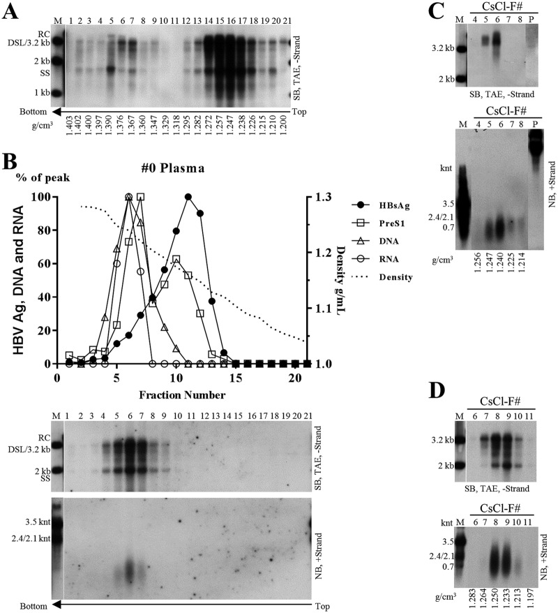
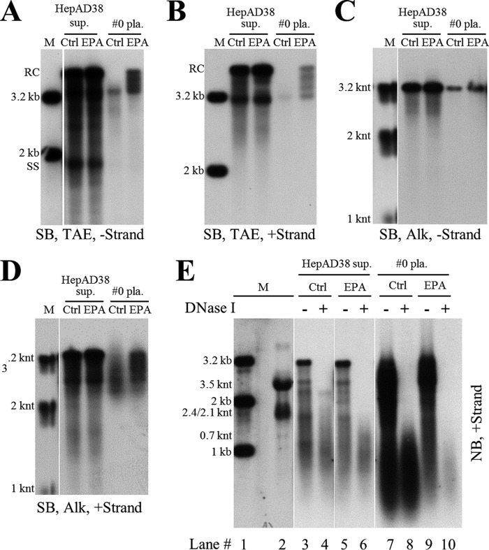
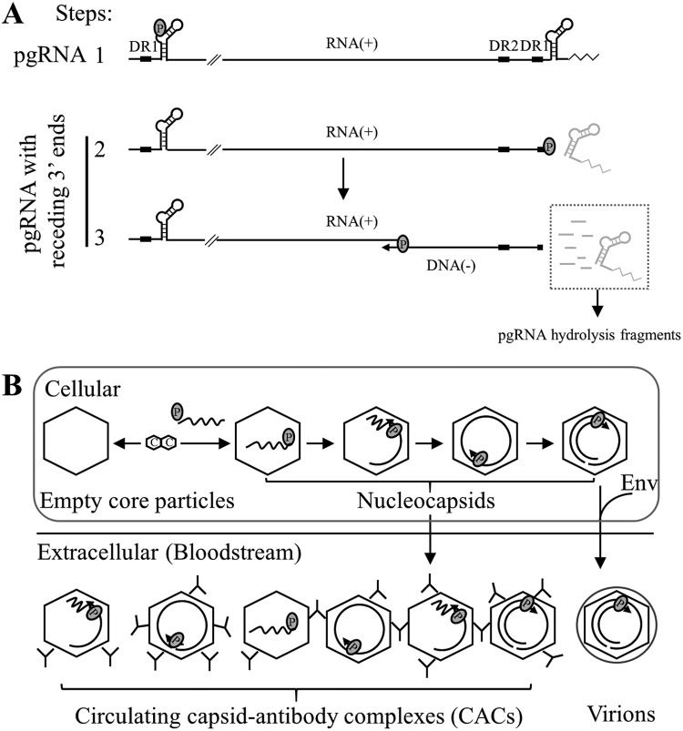
Extracellular HBV RNA has been detected in both HBV-replicating cell culture media and sera from chronic hepatitis B (CHB) patients, but its exact origin and composition remain controversial. Here, we demonstrated that extracellular HBV RNA species were of heterogeneous lengths, ranging from the length of pregenomic RNA to a few hundred nucleotides. In cell models, these RNAs were predominantly associated with naked capsids, although virions also harbored a minority of them. Moreover, HBV RNAs in hepatitis B patients' blood circulation were localized in unenveloped capsids in the form of capsid-antibody complexes (CACs) and in virions. Furthermore, we showed that extracellular HBV RNAs could serve as the template for viral DNA synthesis. In conclusion, extracellular HBV RNAs mainly consist of pgRNA or the pgRNA species degraded by the RNase H domain of the polymerase in the process of viral DNA synthesis and circulate as CACs and virions. Their presence in blood circulation of CHB patients may be exploited to develop novel biomarkers for HBV persistence.IMPORTANCE Although increasing evidence suggests the presence of extracellular HBV RNA species, their origin and molecular forms are still under debate. In addition to the infectious virions, HBV is known to secrete several species of incomplete viral particles, including hepatitis B surface antigen (HBsAg) particles, naked capsids, and empty virions, during its replication cycle. Here, we demonstrated that extracellular HBV RNAs were associated with naked capsids and virions in HepAD38 cells. Interestingly, we found that unenveloped capsids circulate in the blood of hepatitis B patients in the form of CACs and, together with virions, serve as vehicles carrying these RNA molecules. Moreover, extracellular HBV RNAs are heterogeneous in length and represent either pregenomic RNA (pgRNA) or products of incomplete reverse transcription during viral replication. These findings provide a conceptual basis for further application of extracellular RNA species as novel biomarkers for HBV persistence.

Keywords: capsid; capsid-antibody complex; extracellular HBV RNA; hepatitis B virus.


Figures

FIG 1
FIG 1
Sucrose gradient separation and analysis of viral particles from HepAD38 cell culture supernatant. (A) Distribution of hepatitis B viral particle-associated antigens and DNA/RNA in sucrose gradient. Viral particles prepared from HepAD38 cell culture supernatant (via PEG 8000 precipitation) were layered over a 10% to 60% (wt/wt) sucrose gradient for ultracentrifugation separation. Fractions were collected from top to bottom, and HBsAg level was analyzed by enzyme-linked immunosorbent assay (ELISA). HBsAg and viral DNA and RNA (quantified from gray density of bands in panel B) signals and sucrose density were plotted together. Viral particles were first resolved by native agarose gel electrophoresis, followed by immunoblotting (IB) of HBV envelope and core proteins with anti-HBsAg and anti-HBcAg antibodies. (B) Detection of viral DNA/RNA by Southern or Northern blotting. Total viral nucleic acids were extracted by the SDS-proteinase K method, and viral DNA (extracted from one-tenth of the samples used for Northern blotting) and RNA (treated with DNase I) were detected by Southern and Northern blot analyses with minus- or plus-strand-specific riboprobes, respectively. Symbols of HBsAg particles, empty virions (without nucleic acid), virions (with RC DNA), and naked capsids (empty or with nucleic acids) are depicted on the lower right side of panel A. Blank, no nucleic acids; two centered and gapped circles, RC DNA; straight line, SS DNA; wavy lines, pgRNA; M, markers (50 pg of 1-kb, 2-kb, and 3.2-kb DNA fragments released from plasmids as the DNA ladder or total RNA extracted from HepAD38 cells as the RNA ladder).
FIG 2
FIG 2
CsCl density gradient separation and analysis of viral particles from HepAD38 cell culture supernatant. (A) Native agarose gel analysis of viral particles. Culture supernatant of HepAD38 cells was concentrated (via ultrafiltration) and fractionated by CsCl density gradient centrifugation (3 ml of 1.18 g/cm3 CsCl solution in the upper layer and 1.9 ml of 1.33 g/cm3 CsCl solution in the lower layer). Viral particles in each fraction were resolved by native agarose gel electrophoresis, followed by detection of viral antigens with anti-HBsAg and anti-HBcAg antibodies and viral DNA by hybridization with minus-strand-specific riboprobe. (B to F) Southern and Northern blot detection of viral nucleic acids. Viral DNAs were separated by electrophoresis through Tris-acetate-EDTA (TAE) or alkaline (ALK) agarose gel for Southern blotting with minus- or plus-strand-specific riboprobes. Viral RNA was obtained by treatment with total nucleic acids with DNase I and separated by formaldehyde-MOPS agarose gel, followed by Northern blotting. (G) Quantification of viral DNA and RNA in naked capsids or virions. Fractions containing naked capsids (fractions 3 to 7) or virions (fractions 10 to 21) were pooled, and viral DNA and RNA were quantified by PCR. (H) DNA and RNA ratios in naked capsids and virions calculated based on quantitative results. Asterisks indicate unknown high-density viral particles detected by anti-HBcAg or anti-HBsAg antibodies but devoid of any HBV-specific nucleic acids. M, markers (E. coli-derived HBV capsids or DNA and RNA ladders as described in the legend to Fig. 1).
FIG 3
FIG 3
Characterization of HBV DNA and RNA in sera of CHB patients. (A and B) Analyses of serum viral DNA from CHB patients by Southern blotting. Viral DNA was extracted from serum samples obtained from forty-five chronic hepatitis B patients (20% of input sample used for protein A/G agarose beads pulldown) and subjected to Southern blot analysis. Alternatively, these samples were first incubated with protein A/G agarose beads, and then viral DNA in the pulldown mixtures was analyzed by Southern blotting. Serum samples selected for further examining are marked with arrows, and samples with SS DNA detection are labeled with asterisks. (C) Protein A/G agarose bead pulldown of viral particles. Sera (25 μl each) from CHB patients 37, 38, 14, and 35 (M1, mixture one) or from patients 17, 21, 42, and 44 (M2, mixture two) were pooled and incubated with protein A/G agarose beads. Viral DNA in input sera, protein A/G bead pulldown mixtures (beads), and the remaining supernatants (sup.) were extracted and subjected to Southern blot analysis. (D) Northern blot detection of serum viral RNA from patients 37, 38, 14, 35, 17, 21, 42, and 44. Total RNA were extracted from serum samples by TRIzol reagent and treated with DNase I before Northern blot analysis. (E to G) Southern blot analyses of viral DNA from selected samples. Viral DNA was separated by electrophoresis through TAE or alkaline agarose gels, followed by Southern blot detection with the indicated riboprobes.
FIG 4
FIG 4
CsCl density gradient analysis of hepatitis B viral particles. (A and B) CsCl density gradient analysis of viral particles in patient sera. One hundred-microliter volumes of serum mixture from patients 37, 38, 14, and 35 (25 μl each) and 100 μl serum from patient 17 were separated by CsCl density gradient centrifugation (2 ml of 1.18 g/cm3 CsCl solution in the upper layer and 2.9 ml of 1.33 g/cm3 CsCl solution in the lower layer). Viral DNA in each fraction was extracted and detected by Southern blotting. (C to G) CsCl density gradient analysis of viral particles treated with detergent or anti-HBcAg antibody (Ab). Concentrated HepAD38 cell culture supernatant (250 μl each) (via ultrafiltration) was either mixed with anti-HBcAg antibody (10 μl) followed by incubation without (C) or with NP-40 (final concentration, 1%) (D) for 1 h at room temperature and 4 h on ice or treated with only NP-40 (G) and then fractionated by CsCl density gradient ultracentrifugation. Sera from CHB patient 46 either left untreated (E) or treated with NP-40 (final concentration, 1%) (F) were fractionated by CsCl density gradient ultracentrifugation. Viral DNA in each fraction was extracted and subjected to Southern blot analyses.
FIG 5
FIG 5
EM analysis of hepatitis B viral particles. (A and B) EM of E. coli-derived HBV capsids incubated without or with anti-HBcAg antibody. (C) EM of viral particles prepared from sera of CHB patients. Serum mixtures (obtained from patients 11, 22, 23, 27, 28, 30, and 41) depleted of HBsAg particles were negatively stained and examined with an electron microscope. The 42-nm HBV virions (arrowhead) and 27-nm naked capsids (arrow) are indicated, while the smaller 22-nm rods and spheres of HBsAg particles could also be observed but are not pointed out. Scale bars indicate 200 nm or 500 nm.
FIG 6
FIG 6
Native agarose gel analysis of viral particles in sera from hepatitis B patients. (A) Native agarose gel analysis of viral particles from HepAD38 cell culture supernatant. Ten microliters of HepAD38 cell culture supernatant (concentrated by ultrafiltration) incubated with or without anti-HBcAg antibody was resolved by native (TAE) agarose gel (0.8%) electrophoresis, followed by hybridization with minus-strand-specific riboprobe. (B and C) Native agarose gel analysis of viral particles from serum samples of hepatitis B patient in buffer with different pH values. Ten microliters of concentrated HepAD38 cell culture supernatant, plasma sample of patient 0 (not concentrated), and serum of a chronic hepatitis B carrier without liver inflammation (ctrl serum) were loaded into agarose gels prepared in TAE buffer (pH 8.3) (B, left) or Dunn carbonate buffer (10 mM NaCHO3, 3 mM Na2CO3, pH 9.4) (B, right) and separated overnight. Viral particle-associated DNA was detected by hybridization with specific riboprobe. Sera from patients 37, 38, 14, 35, 17, 21, 42, and 44 (10 μl each) were resolved by electrophoresis through 0.7% high-strength agarose (type IV agarose used for pulsed-field gel electrophoresis) gels prepared in TAE (C, left) or carbonate buffer (C, right), followed by probe hybridization.
FIG 7
FIG 7
Characterization of nucleic acid content within viral particles in plasma sample from patient 0. (A) CsCl density gradient analysis of plasma sample. Plasma from patient 0 was added directly with CsCl salt to a concentration of 21% (wt/wt) or 34% (wt/wt). Two milliliters of the 21% CsCl-plasma mixture was underlayered with 2.9 ml 34% CsCl-plasma mixture, followed by ultracentrifugation. Viral DNA from each fraction was extracted and subjected to Southern blot analysis. (B) Sucrose gradient analysis of concentrated plasma sample. Five hundred microliters of concentrated plasma sample (via ultracentrifugation through a 20% sucrose cushion) was fractionated in a 10% to 60% (wt/wt) sucrose gradient. PreS1 and HBsAg levels were determined by ELISA. Viral DNA and RNA were detected by Southern and Northern blotting with minus- or plus-strand-specific riboprobes. HBsAg, PreS1, and viral DNA and RNA (quantified from gray density of viral DNA/RNA bands, middle and lower) signals and sucrose density were plotted together. (C) Analysis of concentrated plasma sample with lower CsCl density gradient centrifugation. Two hundred fifty microliters of concentrated plasma sample was mixed with 2.2 ml TNE buffer and 2.45 ml of 37% (wt/wt) CsCl-TNE buffer (resulting in a homogenous CsCl solution with density of about 1.18 g/cm3), followed by ultracentrifugation. DNA in viral particle pellets (lane P) stuck to the sidewall of centrifugation tubes and was recovered by digesting with SDS-proteinase K solution. Viral DNA and RNA were subjected to Southern and Northern blot analyses. (D) Analysis of concentrated plasma sample with higher level of CsCl density gradient centrifugation. Two hundred fifty microliters of concentrated plasma sample was mixed with 1 ml of TNE buffer and 1.25 ml of 37% (wt/wt) CsCl-TNE buffer and underlayered with 2.4 ml of 27% (wt/wt) (1.25 g/cm3) CsCl-TNE solution, followed by ultracentrifugation. HBV DNA and RNA was detected by Southern and Northern blotting.
FIG 8
FIG 8
Analysis of extracellular HBV DNA and RNA by EPA. (A to D) Southern blot analysis of viral DNA strand elongation after EPA treatment. EPA was carried out employing HepAD38 cell culture supernatant and plasma sample from patient 0. Total nucleic acids were extracted via the SDS-proteinase K method. Viral DNA was separated by electrophoresis in TAE or alkaline agarose gels, followed by Southern blot analysis with minus- or plus-strand-specific riboprobes. (E) Northern blot analysis of viral RNA changed upon EPA treatment. Total viral nucleic acids (lanes 3, 5, 7, and 9) or RNA (treated with DNase I) (lanes 4, 6, 8, and 10) were separated by formaldehyde-MOPS agarose gel electrophoresis and subjected to Northern blotting.
FIG 9
FIG 9
Mapping and identifying 3′ ends of extracellular HBV RNAs. (A) Northern blot detection of extracellular HBV RNAs with various riboprobes. Viral RNA from cytoplasmic (C) nucleocapsids (lanes 2, 5, 8, 11, 14, and 17) or culture supernatant (S) (lanes 3, 6, 9, 12, 15, and 18) of HepAD38 cells was extracted with TRIzol reagent and treated with DNase I before Northern blot analysis with plus-strand-specific riboprobes spanning the HBV genome as indicated. pgRNA was used as a reference, and map coordinates were numbered according to the sequence of the HBV genome (genotype D, accession number AJ344117.1). (B) Identification of 3′ ends of extracellular HBV RNAs. 3′ Ends of extracellular HBV RNAs were identified by the 3′ RACE method using different HBV-specific anchor primers (the same 5′ primers used for generating templates for producing riboprobes used in panel A, lower). Identified 3′ ends were numbered as described above, and numbers in parentheses indicate the amount of clones with the same 3′ ends. The asterisk indicates unknown nucleic acid copurified with intracellular capsid-associated viral RNA by TRIzol reagent. FL, full-length; Cap, 5′ cap of pregenomic RNA; pA, the polyadenylation site; An, poly(A) tail.
FIG 10
FIG 10
Analysis of HBV DNA and RNA change upon entecavir treatment of HepAD38 cells. (A) Change of total cellular HBV RNA level upon entecavir (ETV) treatment. HepAD38 cells were treated with ETV (0.1 μM) for 4 days, and total cellular RNA was analyzed by Northern blotting with ribosomal RNAs serving as loading controls. (B) Change of intracellular nucleocapsid-associated viral RNA (core RNA) and DNA (core DNA) level after ETV treatment. Cytoplasmic core RNA was extracted by the SDS-proteinase K method and analyzed by Northern blotting. Intracellular nucleocapsids were first separated by native agarose gel electrophoresis, and capsid-associated viral DNA (core DNA) was then probed with minus-strand-specific riboprobe. (C to E) Change of extracellular HBV DNA and RNA level upon ETV treatment. Total nucleic acids in HepAD38 cell culture supernatant were extracted and subjected to Southern and Northern blot analyses with specific riboprobes or quantification by PCR. (F to H) CsCl density gradient analysis of viral DNA/RNA level in naked capsids and virions after ETV treatment. HepAD38 cells were left untreated or were treated with ETV, and culture media were concentrated by ultrafiltration, followed by fractionation in CsCl density gradients as described in the legend to Fig. 4. Viral particles in each fraction were separated by native agarose gel electrophoresis, followed by immunoblotting with anti-HBcAg antibody. Viral DNA and RNA were extracted and subjected to Southern or Northern blot analyses.
FIG 11
FIG 11
Models for the content of extracellular HBV RNAs and the formation of circulating CACs. (A) HBV RNA molecules present in the process of DNA synthesis. HBV RNAs are included in the following DNA synthesis steps: 1, encapsidation of full-length pgRNA into NCs; 2, transfer of polymerase-DNA primer to the 3′ DR1 region and initiation of minus-strand DNA synthesis (3′ epsilon loop of pgRNA will be cleaved by RNase H domain of polymerase); 3, elongation of minus-strand DNA. With the extension of minus-strand DNA, pgRNA will be continuously cleaved from the 3′ end, generating pgRNA fragments with receding 3′ ends and pgRNA hydrolysis fragments. (B) Possible forms of circulating CACs. Intracellular NCs with pgRNA or pgRNA fragment and DNA replicative intermediates released into blood circulation of CHB patients are bound with specific antibodies (IgG), forming various forms of CACs.

References

    1. Hellard ME, Chou R, Easterbrook P. 2017. WHO guidelines on testing for hepatitis B and C–meeting targets for testing. BMC Infect Dis 17:703. doi: 10.1186/s12879-017-2765-2. - DOI - PMC - PubMed
    1. Seeger C, Zoulim F, Mason WS. 2007. Hepadnaviruses, p 2977 In Knipe DM, Howley PM (ed), Fields virology, 5th ed, vol 2, Lippincott, Williams & Wilkins, Philadelphia.
    1. Wynne SA, Crowther RA, Leslie AG. 1999. The crystal structure of the human hepatitis B virus capsid. Mol Cell 3:771–780. doi: 10.1016/S1097-2765(01)80009-5. - DOI - PubMed
    1. Crowther RA, Kiselev NA, Bottcher B, Berriman JA, Borisova GP, Ose V, Pumpens P. 1994. Three-dimensional structure of hepatitis B virus core particles determined by electron cryomicroscopy. Cell 77:943–950. doi: 10.1016/0092-8674(94)90142-2. - DOI - PubMed
    1. Summers J, Mason WS. 1982. Replication of the genome of a hepatitis B–like virus by reverse transcription of an RNA intermediate. Cell 29:403–415. doi: 10.1016/0092-8674(82)90157-X. - DOI - PubMed

Publication types

LinkOut - more resources