. 2018 Nov 27;92(24):e01091-18.
doi: 10.1128/JVI.01091-18. Print 2018 Dec 15.

Targeting of the Nasal Mucosa by Japanese Encephalitis Virus for Non-Vector-Borne Transmission

Affiliations

Targeting of the Nasal Mucosa by Japanese Encephalitis Virus for Non-Vector-Borne Transmission

Obdulio García-Nicolás et al. J Virol. .

Abstract

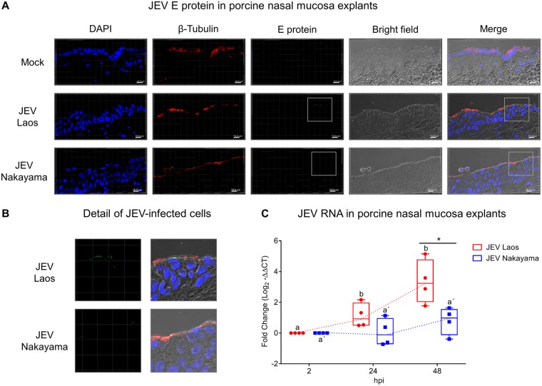
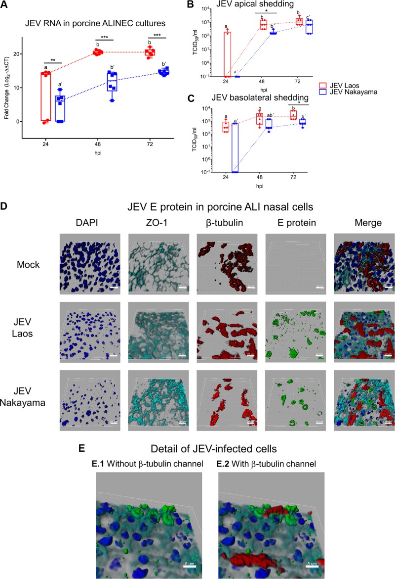
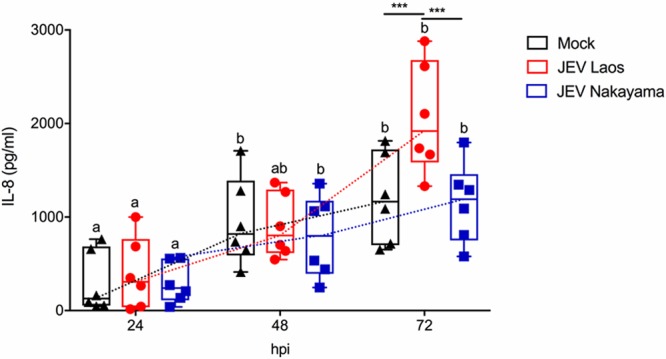
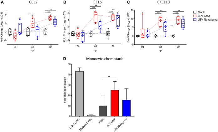
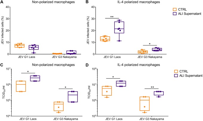
The mosquito-borne Japanese encephalitis virus (JEV) causes severe central nervous system diseases and cycles between Culex mosquitoes and different vertebrates. For JEV and some other flaviviruses, oronasal transmission is described, but the mode of infection is unknown. Using nasal mucosal tissue explants and primary porcine nasal epithelial cells (NEC) at the air-liquid interface (ALI) and macrophages as ex vivo and in vitro models, we determined that the nasal epithelium could represent the route of entry and exit for JEV in pigs. Porcine NEC at the ALI exposed to with JEV resulted in apical and basolateral virus shedding and release of monocyte recruiting chemokines, indicating infection and replication in macrophages. Moreover, macrophages stimulated by alarmins, including interleukin-25, interleukin-33, and thymic stromal lymphopoietin, were more permissive to the JEV infection. Altogether, our data are important to understand the mechanism of non-vector-borne direct transmission of Japanese encephalitis virus in pigs.IMPORTANCE JEV, a main cause of severe viral encephalitis in humans, has a complex ecology composed of a mosquito-waterbird cycle and a cycle involving pigs, which amplifies virus transmission to mosquitoes, leading to increased human cases. JEV can be transmitted between pigs by contact in the absence of arthropod vectors. Moreover, virus or viral RNA is found in oronasal secretions and the nasal epithelium. Using nasal mucosa tissue explants and three-dimensional porcine nasal epithelial cells cultures and macrophages as ex vivo and in vitro models, we determined that the nasal epithelium could be a route of entry as well as exit for the virus. Infection of nasal epithelial cells resulted in apical and basolateral virus shedding and release of monocyte recruiting chemokines and therefore infection and replication in macrophages, which is favored by epithelial-cell-derived cytokines. The results are relevant to understand the mechanism of non-vector-borne direct transmission of JEV.

Keywords: Japanese encephalitis virus; direct contact transmission; macrophages; nasal epithelial cells; pig.


Figures

FIG 1
FIG 1
JEV replicates in porcine nasal explants. Porcine nasal mucosa explants were challenged with JEV Laos or Nakayama at 105 TCID50/sample for 2 h, followed by wash steps and culture in medium for 48 h. In panel A, nuclei were stained with DAPI (blue), cilia with anti-β-tubulin (red), and JEV with anti-E protein 4G2 (green). The scale bar represents 10 µm. Three-dimensional (3D) scans were acquired using confocal microscopy. In panel B, a higher magnification of panel A is shown. In panel C, viral RNA was quantified by real-time RT-PCR. Results were calculated as fold change of the threshold cycle and are represented as 25 to 75% interquartile boxes showing the mean and 95% confidence intervals. Different superscript letters indicate significant difference (P ≤ 0.05) between samples challenged with the same JEV strain (letters without or with apostrophe for JEV Laos and Nakayama strain, respectively); significant differences between distinct JEV strains at the same time postinfection are shown by an asterisk.
FIG 2
FIG 2
JEV infects and replicates in porcine NEC. In panel A, porcine NEC cultures were infected with JEV strains at MOI of 0.1 TCID50/cell, and after 24, 48, and 72 hpi, inserts were collected and viral RNA was quantified by real-time RT-PCR. In panels B and C, viral titers from the apical and basolateral compartments of the same cultures as in panel A are shown. In panels D and E, JEV-infected porcine NEC were analyzed by multicolor immunostaining for nuclei (DAPI, dark blue), cilia (β-tubulin, red), tight junctions (ZO-1; light blue), and JEV E protein. 3D scans were acquired using confocal microscopy; scale bars represent 20 and 8 µm for panels D and E, respectively. The experiment was repeated three times in duplicate. In panels A to C, results are represented as 25 to 75% interquartile boxes showing the means and 95% confidence intervals. Different superscript letters indicate significant differences (P < 0.05) between samples challenged with the same JEV strain (letters without and with apostrophe for the Laos and Nakayama strains, respectively). Significant differences between distinct JEV strains at the same time postinfection are shown by an asterisk for virus titers (A) and for RNA levels (B and C).
FIG 3
FIG 3
JEV-induced cell death in porcine NEC. Porcine NEC were infected with JEV Laos and Nakayama at an MOI of 0.1 TCID50/cell and incubated for 72h. In panel A, dead porcine NEC were labeled with CytoPainter (dark blue) before fixation and then cells were immunolabeled for actin (red) and for JEV E protein (green). In panel B, we immunolabeled cleaved caspase 3 (red), actin (light blue), JEV E protein (green), and nuclei (DAPI, dark blue). CD90-induced apoptosis induction was employed as a positive control. The percentage of E protein expressing dead (A) or apoptotic (B) cells are shown in the upper left corner of each image. 3D scans were acquired using confocal microscopy. The scale bar represents 20 µm. Detailed images of necrotic and apoptotic cells are shown to the far right (A and B).
FIG 4
FIG 4
JEV induces weak proinflammatory responses following infection of porcine NEC. NEC were challenged with JEV Laos and Nakayama at an MOI of 0.1 TCID50/cell. After 24, 48, and 72 hpi, inserts were collected and cytokine gene expression was quantified by RT-qPCR for IL-6 (A), IL-8 (B), IFN-λ3 (C), and SOCS1 (D). The experiment was repeated three times in duplicate. Data are shown as 25% to 75% interquartile boxes with means and 95% confidence intervals. Different superscript letters indicate significant difference (P < 0.05) between samples challenged with the same JEV strain (letters without and with apostrophe for JEV Laos and JEV Nakayama, respectively); differences between distinct JEV strains and the mock control at the same time postinfection are shown by asterisks (*, P ≤ 0.05; **, P ≤ 0.002; ***, P ≤ 0.001).
FIG 5
FIG 5
JEV Laos induces delayed IL-8 secretion following infection of porcine NEC. Porcine NEC were challenged with JEV Laos and Nakayama at an MOI of 0.1 TCID50/cell. After 24, 48, and 72 hpi, basolateral supernatants were collected and IL-8 levels were determined by ELISA. The experiment was repeated three times in duplicate. Data are shown as 25% to 75% interquartile boxes with means and 95% confidence intervals. Different superscript letters indicate significant difference (P < 0.05) between samples challenged with the same JEV strain (letters without and with apostrophe for JEV Laos and JEV Nakayama, respectively); differences between distinct JEV strains and the mock control at the same time postinfection are shown by asterisks (*, P ≤ 0.05; **, P ≤ 0.002; ***, P ≤ 0.001).
FIG 6
FIG 6
JEV-infected porcine NEC express chemokines and induce monocyte chemotaxis. RNA was extracted from NEC and gene expression of chemokines was determined by RT-qPCR for CCL2 (A), CCL5 (B), and CXCL10 (C). Data are shown as 25% to 75% interquartile boxes with means and ±95% confidence intervals. Basolateral medium from porcine NEC at 72 hpi was used for chemotaxis experiments. (D) Mean values ± SD are shown, with different superscript letters indicating significant difference (P ≤ 0.05) between samples challenged with the same JEV strain (letters without and with apostrophe for JEV Laos and JEV Nakayama, respectively); differences between distinct JEV strains and mock at the same time postinfection are shown by asterisks (*, P ≤ 0.05; **, P ≤ 0.002; ***, P ≤ 0.001).
FIG 7
FIG 7
Porcine NEC cultures release factors enhancing porcine macrophage infection by JEV. Basolateral supernatants from NEC at 72 hpi were used to infect nonpolarized and IL-4-polarized macrophages. The percentages of infected cells are shown for nonpolarized (A) and IL-4-polarized macrophages (B). Panels C and D show the virus shed from nonpolarized and IL-4-polarized macrophages, respectively. Results represent those from three independent experiments performed in duplicate. Data are shown as 25% to 75% interquartile boxes with means and 95% confidence intervals. Different superscript letters indicate significant difference (P < 0.05) between samples challenged with the same JEV strain (letters without and with apostrophe for JEV Laos and JEV Nakayama, respectively); differences between distinct JEV strains and the mock control at the same time postinfection are shown by asterisks (*, P ≤ 0.05; **, P ≤ 0.002).
FIG 8
FIG 8
JEV infection is enhanced in IL-4-treated MDM. Porcine MDM were infected with JEV Laos obtained from virus stocks or from the basolateral medium of porcine NEC cultures infected with JEV Laos or Nakayama for 72 h at an MOI of 1 TCID50/cell. After 24 h, E expressing cells were determined by flow cytometry and virus titers in the supernatants measured for JEV Laos and Nakayama (A and B, respectively). All experiments were repeated three independent times in duplicate, and data are represented as 25% to 75% interquartile boxes showing the means and 95% confidence intervals. Significant differences between nonpolarized and IL-4 polarized MDM for the same challenge condition are shown by asterisks (*, P ≤ 0.05; **, P ≤ 0.002).
FIG 9
FIG 9
IL-25, IL-33, and TSLP can enhance infection of MDM. Nonpolarized (A) and IL-4-polarized (B) porcine macrophages were challenged with JEV Laos at an MOI of 1 TCID50/cell. The influence of IL-25, IL-33, and TSLP (all at 10 ng/ml) or combinations thereof was tested when added with the virus inoculum, incubated for 1.5 h, and washed off; after 24 hpi, cells were harvested and E protein-positive cells acquired by flow cytometry. Data represent those from three independent experiments carried out in triplicate and illustrate 25% to 75% interquartile boxes showing the means and 95% confidence intervals. Differences between JEV infection of cells in the presence or absence of the cytokines are shown by asterisks (*, P ≤ 0.05; **, P ≤ 0.002; ***, P ≤ 0.001).
FIG 10
FIG 10
Targeting of the nasal mucosa by JEV. Shown is a graphical summary of the interaction of JEV with NEC and myeloid cells.

References

    1. WHO. 2015. Japanese encephalitis. WHO, Geneva, Switzerland.
    1. Impoinvil DE, Baylis M, Solomon T. 2013. Japanese encephalitis: on the One Health agenda. Curr Top Microbiol Immunol 365:205–247. doi:10.1007/82_2012_243. - DOI - PubMed
    1. Le Flohic G, Porphyre V, Barbazan P, Gonzalez J-P. 2013. Review of climate, landscape, and viral genetics as drivers of the Japanese encephalitis virus ecology. PLoS Negl Trop Dis 7:e2208. doi:10.1371/journal.pntd.0002208. - DOI - PMC - PubMed
    1. Scherer WF, Buescher EL. 1959. Ecologic studies of Japanese encephalitis virus in Japan. Am J Trop Med Hyg 8:644–650. doi:10.4269/ajtmh.1959.8.644. - DOI - PubMed
    1. Weaver SC, Barrett ADT. 2004. Transmission cycles, host range, evolution and emergence of arboviral disease. Nat Rev Microbiol 2:789–801. doi:10.1038/nrmicro1006. - DOI - PMC - PubMed

Publication types

MeSH terms