. 2018 Oct 30;91(18):e1677-e1689.
doi: 10.1212/WNL.0000000000006435. Epub 2018 Oct 3.

Paraneoplastic neuronal intermediate filament autoimmunity

Affiliations

Paraneoplastic neuronal intermediate filament autoimmunity

Eati Basal et al. Neurology. .

Abstract

Objective: To describe paraneoplastic neuronal intermediate filament (NIF) autoimmunity.

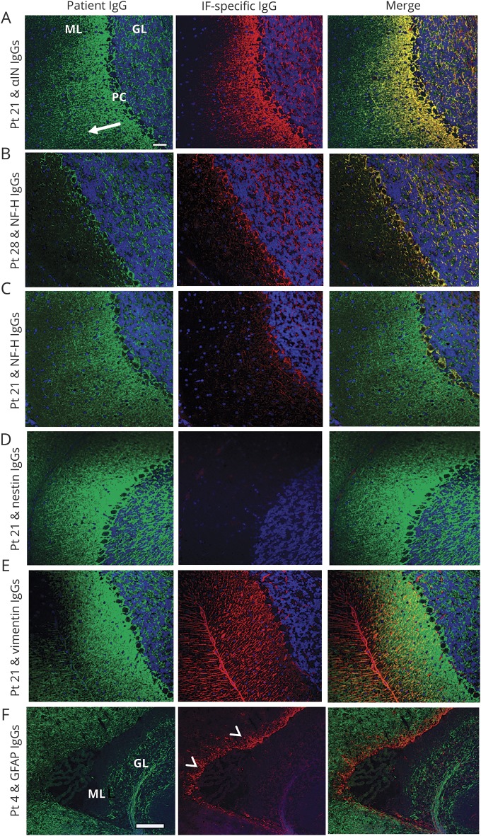
Methods: Archived patient and control serum and CSF specimens were evaluated by tissue-based indirect immunofluorescence assay (IFA). Autoantigens were identified by Western blot and mass spectrometry. NIF specificity was confirmed by dual tissue section staining and 5 recombinant NIF-specific HEK293 cell-based assays (CBAs, for α-internexin, neurofilament light [NfL], neurofilament medium, or neurofilament heavy chain, and peripherin). NIF-immunoglobulin Gs (IgGs) were correlated with neurologic syndromes and cancers.

Results: Among 65 patients, NIF-IgG-positive by IFA and CBAs, 33 were female (51%). Median symptom onset age was 62 years (range 18-88). Patients fell into 2 groups, defined by the presence of NfL-IgG (21 patients, who mostly had ≥4 NIF-IgGs detected) or its absence (44 patients, who mostly had ≤2 NIF-IgGs detected). Among NfL-IgG-positive patients, 19/21 had ≥1 subacute onset CNS disorders: cerebellar ataxia (11), encephalopathy (11), or myelopathy (2). Cancers were detected in 16 of 21 patients (77%): carcinomas of neuroendocrine lineage (10) being most common (small cell [5], Merkel cell [3], other neuroendocrine [2]). Two of 257 controls (0.8%, both with small cell carcinoma) were positive by both IFA and CBA. Five of 7 patients with immunotherapy data improved. By comparison, the 44 NfL-IgG-negative patients had findings of unclear significance: diverse nervous system disorders (p = 0.006), as well as limited (p = 0.003) and more diverse (p < 0.0001) cancer accompaniments.

Conclusions: NIF-IgG detection by IFA, with confirmatory CBA testing that yields a profile including NfL-IgG, defines a paraneoplastic CNS disorder (usually ataxia or encephalopathy) accompanying neuroendocrine lineage neoplasia.


Figures

Figure 1
Figure 1. Algorithm for antigen characterization and 2-step algorithm for the serologic diagnosis of neuronal intermediate filament (NIF) autoimmunity
Algorithm for (A) antigen characterization and (B) 2-step algorithm for the serologic diagnosis of NIF autoimmunity. (B) Each row represents 1 specimen from 65 patients (48 sera, 19 CSF) or controls (237 sera, 20 CSF), all tested by both tissue-based immunofluorescence assay (IFA) and all 5 NIF–immunoglobulin G (IgG) cell-based assays (CBAs). Only 2 controls (both with cancer) were IFA- and CBA-positive. Specificity assurance requires positivity by both IFA plus one or more recombinant NIF CBAs. NfL = neurofilament light chain.
Figure 2
Figure 2. Immunofluorescence patterns of patient immunoglobulin G (IgG) binding to mouse tissues.
Cerebellum (A, E), hippocampus (B, F), and gastric neuronal ganglia and nerves (C, G) exposed to serum of patient 21 (A.a–C.a) and patient 28 (E.a–G.a) or to IgGs affinity-purified from serum of those patients by acid elution from replicas of Western blotted bands (A.b–C.b [65 kDa] and E2–G2 [200 kDa]). Smooth muscle antibody in patient 21 serum partially obscures the neural staining in C.a but not C.b. For comparison, cerebellar staining by commercial α internexin IgG (D) and neurofilament heavy chain IgG (H) are demonstrated (see also figure e-1). Scale bar = 50 μm.
Figure 3
Figure 3. Dual immunostaining of mouse cerebellum with patient immunoglobulin G (IgG) and IgG specific for neuronal or astrocytic intermediate filaments (IF)
Patient IgG (Pt, green) binding to mouse cerebellar cortex colocalizes with commercial IgGs (red) specific for αinternexin (αIN) IgG or neurofilament heavy (NfH) IgG (yellow in merge), but not with nestin, vimentin, or glial fibrillary acidic protein (GFAP). (A) Patient 21 serum (pattern 1) yields a filamentous pattern in the molecular layer (ML), Purkinje cell layer (PC), and granular layer (GL). Staining, most intense in ML and gradually fading from deep to superficial regions (arrow), colocalizes with αIN IgG. (B) Patient 28 serum (pattern 2) yields a staining pattern mostly restricted to the GL and PC layer, and colocalizes with NfH IgG. (C) Patient 21 serum partially colocalizes with NfH IgG, but not with early developmental neuronal intermediate filaments (nestin [D], vimentin [E]). Patient 4 serum (pattern 1) does not colocalize with GFAP (F) which, characteristically, is most prominent in the subventricular zone (arrowheads; the choroid plexus is nonstained). Scale bar = 20 μm except for F = 100 μm.
Figure 4
Figure 4. Western blot characterization of autoantibodies
(A) Rat spinal cord proteins, reduced, denatured, and separated electrophoretically, were probed with commercial neuronal intermediate filament (NIF) immunoglobulin G (IgG) (lanes 1–4), patient IgG (patients 1, 2, 12, 13, and 17 are in lanes 6–10, respectively), or healthy control IgG (lanes 12–16). Lanes 5 and 11 are empty. Patient IgGs bind to 2 or more prominent bands (molecular weight 65 kDa, 70 kDa, 150 kDa, or 200 kDa), consistent with α internexin (αIN), neurofilament light chain (NfL), neurofilament medium chain (NfM), and neurofilament heavy chain (NfH). (B) Proteins from rat spinal cord lysate bound by patient IgGs (12 [left] and 17 [right]) and immunoprecipitated by adsorption to protein G-complexed magnetic beads were separated electrophoretically and subjected to Western blot. Probing with 4 commercial IgGs specific for NfH, NfM, NfL, and αIN revealed bands with anticipated molecular weights for those NIF proteins. The corresponding proteins were analyzed by mass spectrometry.
Figure 5
Figure 5. Patient immunoglobulin G (IgG) binding to HEK-293 cells transfected with cDNAs encoding green-fluorescent protein (GFP)–tagged human neuronal intermediate filaments (NIFs)
Patient IgGs (red) had diverse NIF reactivities. Illustrative examples include (A) patient 2 serum bound to α internexin (αIN), neurofilament light chain (NfL), neurofilament medium chain (NfM), neurofilament heavy chain (NfH), and peripherin; (B) patient 22 serum bound solely to αIN; (C) patient 32 serum bound to NfM only; and (D) patient 28 serum bound to NfH only. Scale bar = 20 μm.
Figure 6
Figure 6. Neuronal intermediate filament (NIF) expression in metastatic Merkel cell carcinoma
Metastatic tumor cells in lymph node of patient 8 (serum immunoglobulin G [IgG] positive for all NIFs except peripherin) show foci of cytokeratin immunoreactivities, AE1/AE3 (A) and CK20 (B), and universal synaptophysin immunoreactivity (C), consistent with Merkel cell carcinoma. Additional immunoreactivities demonstrated: α internexin (αIN; D), neurofilament light chain (NfL; E), neurofilament medium chain (NfM; F), and neurofilament heavy chain (NfH; G); peripherin immunoreactivity was lacking (H). Scale bar = 20 μm.

References

    1. McKeon A, Pittock SJ. Paraneoplastic encephalomyelopathies: pathology and mechanisms. Acta Neuropathol 2011;122:381–400. - PubMed
    1. Dalmau J, Gleichman AJ, Hughes EG, et al. Anti-NMDA-receptor encephalitis: case series and analysis of the effects of antibodies. Lancet Neurol 2008;7:1091–1098. - PMC - PubMed
    1. Fang B, McKeon A, Hinson SR, et al. Autoimmune glial fibrillary acidic protein astrocytopathy: a novel meningoencephalomyelitis. JAMA Neurol 2016;73:1297–1307. - PubMed
    1. Flanagan EP, Hinson SR, Lennon VA, et al. Glial fibrillary acidic protein immunoglobulin G as biomarker of autoimmune astrocytopathy: analysis of 102 patients. Ann Neurol 2017;81:298–309. - PubMed
    1. Braxton DB, Williams M, Kamali D, Chin S, Liem R, Latov N. Specificity of human anti-neurofilament autoantibodies. J Neuroimmunol 1989;21:193–203. - PubMed