. 2018 Nov 23;293(47):18378-18386.
doi: 10.1074/jbc.RA118.004202. Epub 2018 Oct 3.

A helical LC3-interacting region mediates the interaction between the retroviral restriction factor Trim5α and mammalian autophagy-related ATG8 proteins

Affiliations

A helical LC3-interacting region mediates the interaction between the retroviral restriction factor Trim5α and mammalian autophagy-related ATG8 proteins

Jeremy R Keown et al. J Biol Chem. .

Abstract

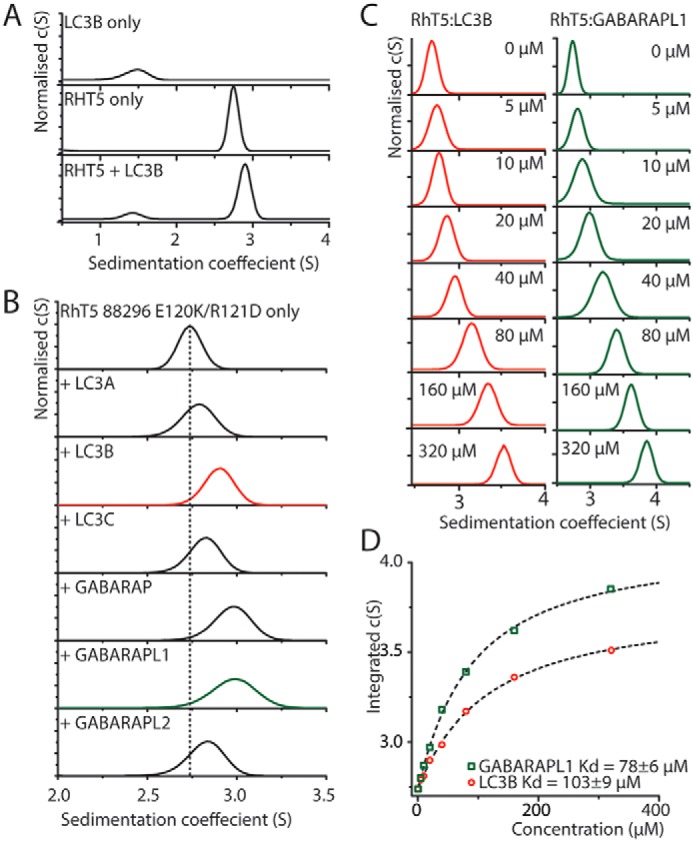
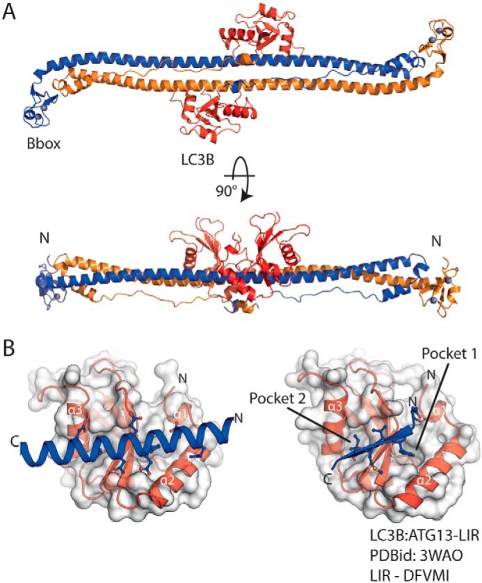
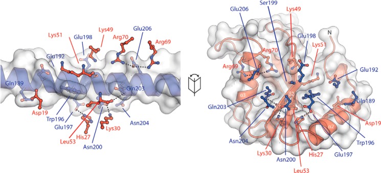
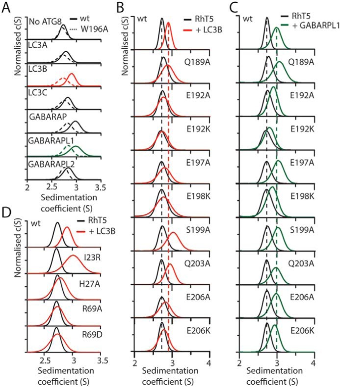
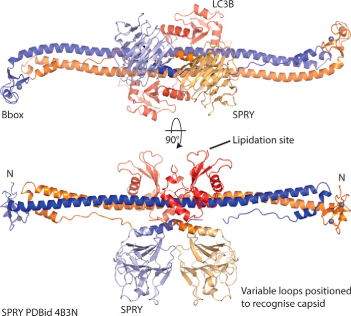
The retroviral restriction factor tripartite motif-containing 5α (Trim5α) acts during the early postentry stages of the retroviral life cycle to block infection by a broad range of retroviruses, disrupting reverse transcription and integration. The mechanism of this restriction is poorly understood, but it has recently been suggested to involve recruitment of components of the autophagy machinery, including members of the mammalian autophagy-related 8 (ATG8) family involved in targeting proteins to the autophagosome. To better understand the molecular details of this interaction, here we utilized analytical ultracentrifugation to characterize the binding of six ATG8 isoforms and determined the crystal structure of the Trim5α Bbox coiled-coil region in complex with one member of the mammalian ATG8 proteins, autophagy-related protein LC3 B (LC3B). We found that Trim5α binds all mammalian ATG8s and that, unlike the typical LC3-interacting region (LIR) that binds to mammalian ATG8s through a β-strand motif comprising approximately six residues, LC3B binds to Trim5α via the α-helical coiled-coil region. The orientation of the structure demonstrated that LC3B could be accommodated within a Trim5α assembly that can bind the retroviral capsid. However, mutation of the binding interface does not affect retroviral restriction. Comparison of the typical linear β-strand LIR with our atypical helical LIR reveals a conservation of the presentation of residues that are required for the interaction with LC3B. This observation expands the range of LC3B-binding proteins to include helical binding motifs and demonstrates a link between Trim5α and components of the autophagosome.

Keywords: LC3B; LIR; Trim5alpha; antiviral protein; autophagy; human immunodeficiency virus (HIV); protein complex; protein-protein interaction; retroviral restriction factor; retrovirus.


Conflict of interest statement

The authors declare that they have no conflicts of interest with the contents of this article

Figures

Figure 1.
Figure 1.
SV–AUC analysis shows that purified Trim5α binds to six isoforms of mATG8s. A, c(s) analysis of SV–AUC of 20 μm LC3B, RhT5 88–296 EK/RD, or an equimolar mixture. B, c(s) analysis of 20 μm of RhT5 88–296 EK/RD and equimolar concentration of the six mATG8 isoforms. C, c(s) analysis of 20 μm RhT5 88–296 EK/RD and increasing concentrations (0–320 μm) of either LC3B or GABARAPL1. D, peak centroid position derived from integration of the c(s) function from C versus LC3B or GABARAPL1 concentration. A one-site binding model has been used to determine the equilibrium dissociation constant (dashed lines).
Figure 2.
Figure 2.
The coiled coil of Trim5α binds LC3B through a helical motif. A, cartoon representation of the RhT5 88–296 EK/RD–LC3B protein complex. Chain A/B (blue/orange) form the Trim5α antiparallel coiled coil dimer, LC3B molecules are red, and zinc atoms are shown as spheres (silver). B, expanded view of the LC3B-binding site (left) and comparison with a typical β-strand LIR motif (LC3B–ATG13-LIR PDB code 3WAO). Both the helical and β-strand LIR occupy and proceed in the same orientation through the LC3B-binding site.
Figure 3.
Figure 3.
Interactions mediating binding of LC3B to the Trim5α coiled coil. A fold out of the LC3B–RhT5α interaction with RhT5α in blue and LC3B in red is shown. Residues from the opposing molecule are shown as ball-and-stick representation with potential hydrogen bonds as dashed lines. Trp196 of Trim5α occupies pocket 1 of LC3B.
Figure 4.
Figure 4.
Mutational analysis of the LC3B-RhT5α interface. A, c(s) analysis of the RhT5 88–296 EK/RD-mATG8 interaction with all mATG8 proteins at 20 μm equimolar concentration, WT (solid lines), and W196A RhT5 88–296 EK/RD (dashed lines). B and C, c(s) analysis of mixtures of WT and mutant RhT5 88–296 EK/RD with either LC3B (B) or GABARAPL1 (C). Dashed lines indicate the position of RhT5 88–296 EK/RD (black) or with the addition of either LC3B (red) or GABARAPL1 (green). D, c(s) analysis of LC3B mutants (red) mixed with WT RhT5 88–296 EK/RD (black).
Figure 5.
Figure 5.
Model of Trim5α–LC3B complex. The SPRY domains from Rhesus Trim5α (PDB code 4B3N) are positioned on the Trim5α Bbox coiled coil (PDB code 4TN3) by superposition of common residues. The structure of the RhT5α–LC3B complex (this work) was then superimposed on 4TN3 to position the LC3B molecules. This model positions the variable loops of the SPRY domain to recognize capsid, whereas the lipidation site at the C terminus of LC3B is available to be incorporated in the autophagosomal membrane.

References

    1. Nakatogawa H., Ichimura Y., and Ohsumi Y. (2007) Atg8, a ubiquitin-like protein required for autophagosome formation, mediates membrane tethering and hemifusion. Cell 130, 165–178 10.1016/j.cell.2007.05.021 - DOI - PubMed
    1. Popelka H., and Klionsky D. J. (2015) Analysis of the native conformation of the LIR/AIM motif in the Atg8/LC3/GABARAP-binding proteins. Autophagy 11, 2153–2159 10.1080/15548627.2015.1111503 - DOI - PMC - PubMed
    1. Birgisdottir Å. B., Lamark T., and Johansen T. (2013) The LIR motif: crucial for selective autophagy. J. Cell Sci. 126, 3237–3247 - PubMed
    1. Suzuki H., Tabata K., Morita E., Kawasaki M., Kato R., Dobson R. C., Yoshimori T., and Wakatsuki S. (2014) Structural basis of the autophagy-related LC3/Atg13 LIR complex: recognition and interaction mechanism. Structure 22, 47–58 10.1016/j.str.2013.09.023 - DOI - PubMed
    1. McEwan D. G., Popovic D., Gubas A., Terawaki S., Suzuki H., Stadel D., Coxon F. P., Miranda de Stegmann D., Bhogaraju S., Maddi K., Gatti E., Helfrich M. H., Wakatsuki S., Behrends C., Pierre P., et al. (2015) PLEKHM1 regulates autophagosome-lysosome fusion through HOPS complex and LC3/GABARAP proteins. Mol. Cell 57, 39–54 10.1016/j.molcel.2014.11.006 - DOI - PubMed

Publication types

MeSH terms