. 2018 Oct 3;10(4):175.
doi: 10.3390/pharmaceutics10040175.

Combining Microbubble Contrast Agent with Pulsed-Laser Irradiation for Transdermal Drug Delivery

Affiliations

Combining Microbubble Contrast Agent with Pulsed-Laser Irradiation for Transdermal Drug Delivery

Ai-Ho Liao et al. Pharmaceutics. .

Abstract

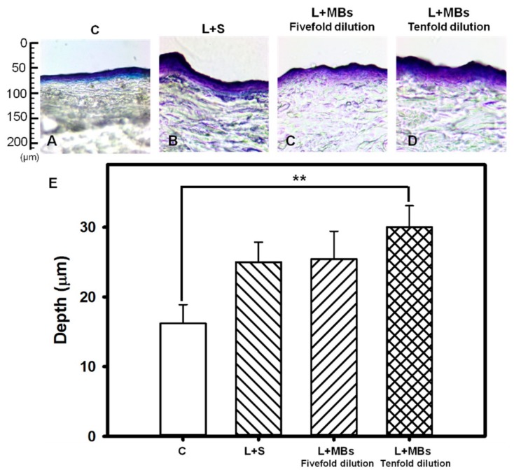
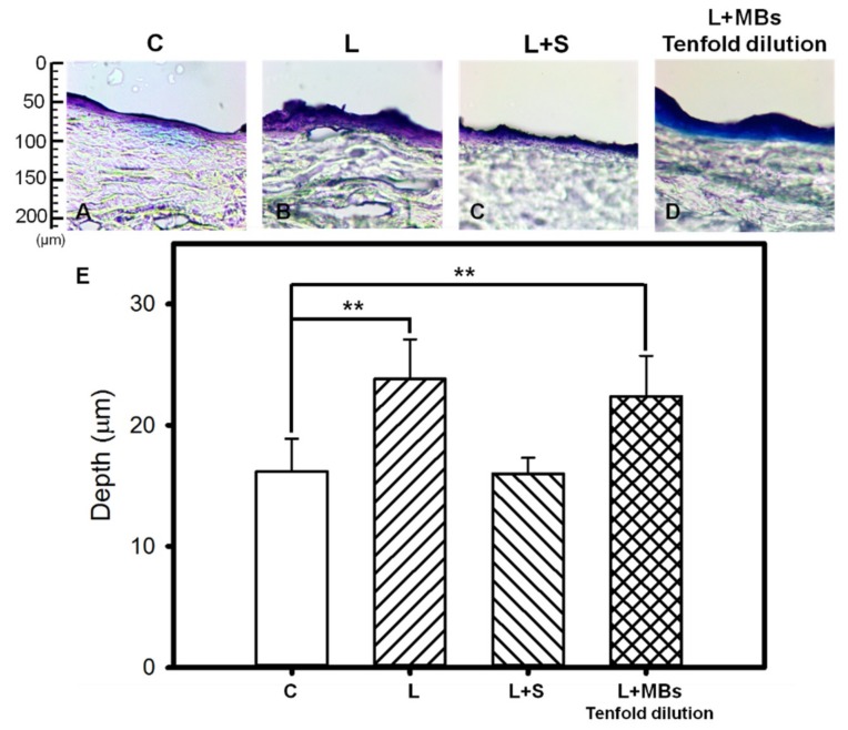
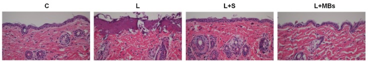
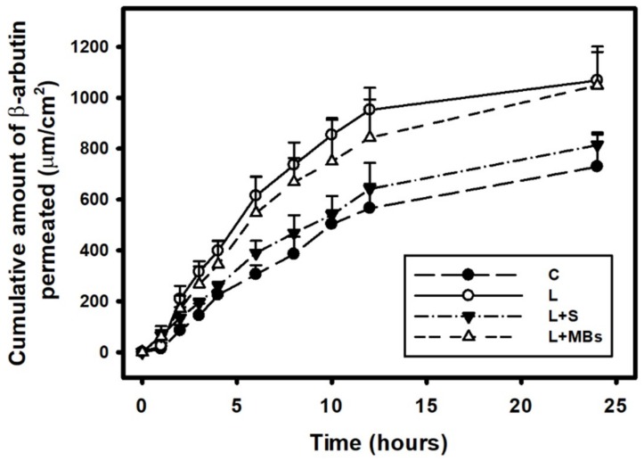
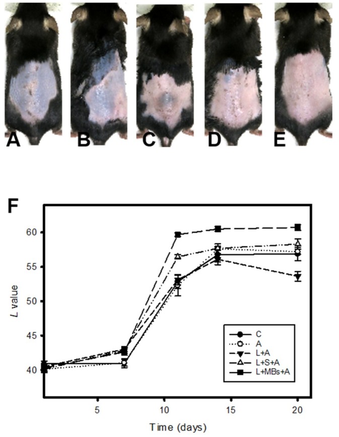
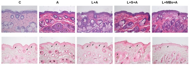
The optodynamic process of laser-induced microbubble (MB) cavitation in liquids is utilized in various medical applications. However, how incident laser radiation interacts with MBs as an ultrasound contrast agent is rarely estimated when the liquid already contains stable MBs. The present study investigated the efficacy of the laser-mediated cavitation of albumin-shelled MBs in enhancing transdermal drug delivery. Different types and conditions of laser-mediated inertial cavitation of MBs were first evaluated. A CO₂ fractional pulsed laser was selected for combining with MBs in the in vitro and in vivo experiments. The in vitro skin penetration by β-arbutin after 2 h was 2 times greater in the group combining a laser with MBs than in the control group. In small-animal experiments, the whitening effect on the skin of C57BL/6J mice in the group combining a laser with MBs on the skin plus penetrating β-arbutin increased (significantly) by 48.0% at day 11 and 50.0% at day 14, and then tended to stabilize for the remainder of the 20-day experimental period. The present results indicate that combining a CO₂ laser with albumin-shelled MBs can increase skin permeability so as to enhance the delivery of β-arbutin to inhibit melanogenesis in mice without damaging the skin.

Keywords: arbutin; cavitation; laser; transdermal; ultrasound contrast agents.


Conflict of interest statement

The authors declare that they have no conflict of interest.

Figures

Figure 1
Figure 1
A Nd:YAG laser setup for measuring laser-induced microbubble (MB) disruption.
Figure 2
Figure 2
Microscopy images (two leftmost columns) of fivefold-diluted MBs without and with continuous- and pulsed-laser irradiation at 60, 120, and 180 s. The controls labeled as before and after refer to the original (fivefold-diluted) MBs and the MBs remaining on the slide after 180 s in the absence of laser irradiation. The microscopy images in the two rightmost columns are the images of the corresponding leftmost columns converted into grayscale images.
Figure 3
Figure 3
Microscopy images of five- and tenfold-diluted MBs without and with Nd:YAG pulsed-laser irradiation at 60, 120, and 180 s.
Figure 4
Figure 4
Microscopy images of tenfold-diluted MBs without and with irradiation by a clinical CO2 fractional pulsed laser one, three, and seven times.
Figure 5
Figure 5
Light-microscope evaluation of pigskin samples with no treatment (group C) (A); for irradiation by the Nd:YAG pulsed laser combined with saline (group L + S) (B); and for laser irradiation combined with MBs (group L + MBs) diluted fivefold (C) and tenfold (D); (E) Quantification of the penetration depths of Evans blue in panels (AD) (** p < 0.01). Data are mean and SD values.
Figure 6
Figure 6
Light-microscope evaluation of pigskin samples in group C (A); for irradiation by the CO2 fractional pulsed laser directly (group L) (B); and for laser irradiation combined with saline (group L + S) (C) and tenfold-diluted MBs (group L + MBs) (D); (E) Quantification of the penetration depths of Evans blue in panels (AD) (** p < 0.01). Data are mean and SD values.
Figure 7
Figure 7
Light-microscope hematoxylin and eosin (HE) evaluation of pigskin samples in group C, for irradiation with the CO2 fractional pulsed laser directly (group L), and for laser irradiation combined with saline (group L + S) and tenfold-diluted MBs (group L + MBs).
Figure 8
Figure 8
In vitro drug penetration in the different experimental groups (see Table 2) through pigskin in a Franz diffusion cell at 36–37 °C. Data are mean and SD values.
Figure 9
Figure 9
Photographs of mouse skin in group C (A); for the application of penetrating β-arbutin alone (group A) (B); for the laser irradiating the skin directly with the application of penetrating β-arbutin (group L + A) (C); for the laser irradiating the skin covered by saline and with the application of penetrating β-arbutin (group L + S + A) (D); and for the laser irradiating the skin combined with MBs on the skin and with the application of penetrating β-arbutin (group L + MBs + A) (E) over a 20-day treatment period; (F) Quantification of the skin-whitening effects of β-arbutin on ultraviolet-radiation-induced hyperpigmentation for the groups in panels (AE). Data are mean and SD values.
Figure 10
Figure 10
Histology images (HE, (upper row); Fontana-Masson silver nitrate, (lower row)) for groups C, A, L + A, L + S + A, and L + MBs + A at day 20. Arrows indicate melanocytes in the basal layer of the epidermis.

Similar articles

Cited by

References

    1. Tzanakis I., Lebon G.S., Eskin D.G., Pericleous K.A. Characterizing the cavitation development and acoustic spectrum in various liquids. Ultrason. Sonochem. 2017;34:651–662. doi: 10.1016/j.ultsonch.2016.06.034. - DOI - PubMed
    1. Dalecki D. Biological Effects of Microbubble-Based Ultrasound Contrast Agents. In: Emilio Q., editor. Contrast Media in Ultrasonography: Basic Principles and Clinical Applications. Springer-Verlag; Berlin/Heidelberg, Germany: 2005. pp. 77–85.
    1. Rota C., Raeman C.H., Child S.Z., Dalecki D. Detection of acoustic cavitation in the heart with microbubble contrast agents in vivo: A mechanism for ultrasound-induced arrhythmias. J. Acoust. Soc. Am. 2006;120:2958–2964. doi: 10.1121/1.2346132. - DOI - PubMed
    1. Van der Wouw P.A., Brauns A.C., Bailey S.E., Powers J.E., Wilde A.A. Premature ventricular contractions during triggered imaging with ultrasound contrast. J. Am. Soc. Echocardiogr. 2000;13:288–294. doi: 10.1067/mje.2000.103865. - DOI - PubMed
    1. Li P., Cao L.Q., Dou C.Y., Armstrong W.F., Miller D. Impact of myocardial contrast echocardiography on vascular permeability: An in vivo dose response study of delivery mode, pressure amplitude and contrast dose. Ultrasound Med. Biol. 2003;29:1341–1349. doi: 10.1016/S0301-5629(03)00988-8. - DOI - PubMed

LinkOut - more resources