

CLIC1 and CLIC4 complement CA125 as a diagnostic biomarker panel for all subtypes of epithelial ovarian cancer
- PMID: 30282979
- PMCID: PMC6170428
- DOI: 10.1038/s41598-018-32885-2
CLIC1 and CLIC4 complement CA125 as a diagnostic biomarker panel for all subtypes of epithelial ovarian cancer
Abstract
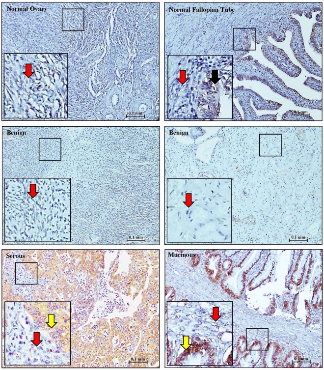
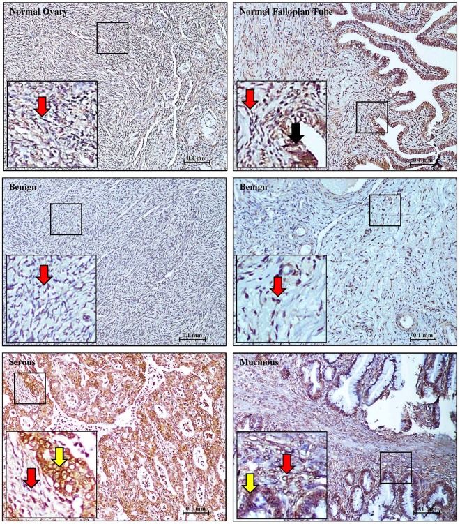
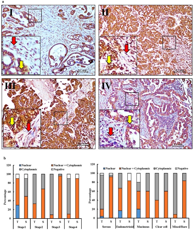
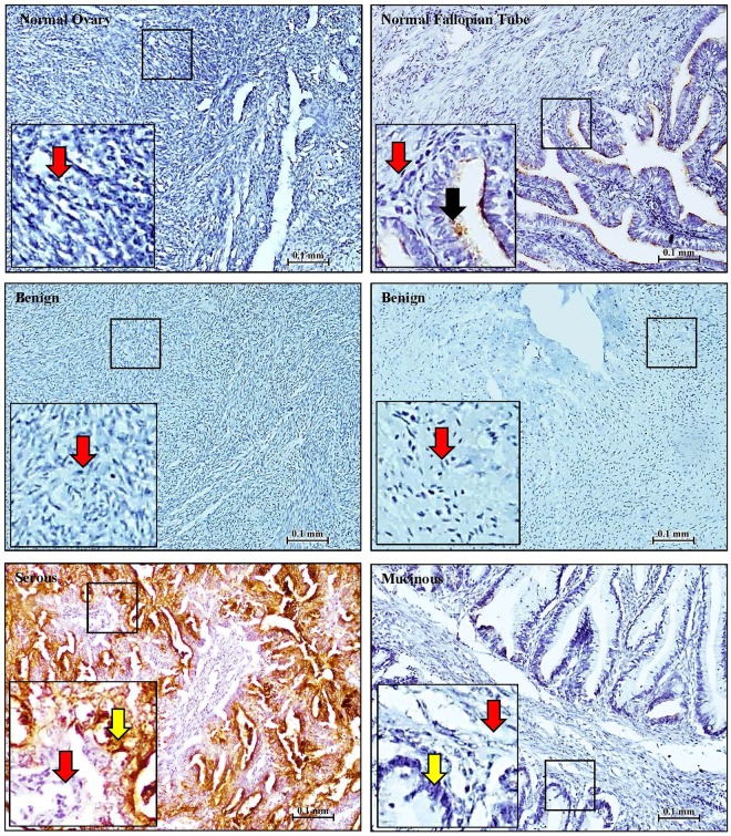
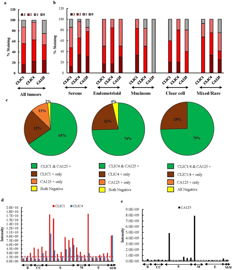
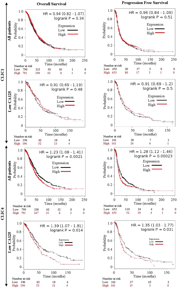
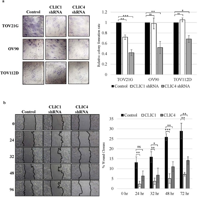
New plasma and tissue biomarkers of epithelial ovarian cancer (EOC) could improve early diagnosis and post-diagnosis clinical management. Here we investigated tissue staining and tissue secretion of CLIC1 and CLIC4 across EOC subtypes. CLIC1 and CLIC4 are two promising biomarkers we previously showed were elevated in EOC patient sera. Individually, CLIC1 or CLIC4 stained larger percentages of malignant tumors across all EOC subtypes compared with CA125, particularly early stage and mucinous tumors. CLIC4 also stained benign tumors but staining was limited to nuclei; whereas malignant tumors showed diffuse cellular staining of stromal and tumor cells. Both proteins were shed by all EOC subtypes tumors in short term organ culture at more consistent levels than CA125, supporting their potential as pan-subtype serum and tissue biomarkers. Elevated CLIC4 expression, but not CLIC1 expression, was a negative indicator of patient survival, and CLIC4 knockdown in cultured cells decreased cell proliferation and migration indicating a potential role in tumor progression. These results suggest CLIC1 and CLIC4 are promising serum and tissue biomarkers as well as potential therapeutic targets for all EOC subtypes. This justifies development of high throughput serum/plasma biomarker assays to evaluate utility of a biomarker panel consisting of CLIC1, CLIC4 and CA125.
Conflict of interest statement
The authors declare no competing interests.
Figures
Similar articles
-
Serum CA125 in combination with ferritin improves diagnostic accuracy for epithelial ovarian cancer.Br J Biomed Sci. 2018 Apr;75(2):66-70. doi: 10.1080/09674845.2017.1394051. Epub 2018 Feb 16. Br J Biomed Sci. 2018. PMID: 29452533
-
Tissue CA125 and HE4 Gene Expression Levels Offer Superior Accuracy in Discriminating Benign from Malignant Pelvic Masses.Asian Pac J Cancer Prev. 2016;17(1):323-33. doi: 10.7314/apjcp.2016.17.1.323. Asian Pac J Cancer Prev. 2016. PMID: 26838232
-
Diagnostic Model of Serum miR-193a-5p, HE4 and CA125 Improves the Diagnostic Efficacy of Epithelium Ovarian Cancer.Pathol Oncol Res. 2018 Oct;24(4):739-744. doi: 10.1007/s12253-018-0392-x. Epub 2018 Mar 8. Pathol Oncol Res. 2018. PMID: 29520570
-
Chloride channels in cancer: Focus on chloride intracellular channel 1 and 4 (CLIC1 AND CLIC4) proteins in tumor development and as novel therapeutic targets.Biochim Biophys Acta. 2015 Oct;1848(10 Pt B):2523-31. doi: 10.1016/j.bbamem.2014.12.012. Epub 2014 Dec 26. Biochim Biophys Acta. 2015. PMID: 25546839 Review.
-
[Diagnostic and prognostic value of tumor markers, scores (clinical and biological) algorithms, in front of an ovarian mass suspected of an epithelial ovarian cancer: Article drafted from the French Guidelines in oncology entitled "Initial management of patients with epithelial ovarian cancer" developed by FRANCOGYN, CNGOF, SFOG, GINECO-ARCAGY under the aegis of CNGOF and endorsed by INCa].Gynecol Obstet Fertil Senol. 2019 Feb;47(2):134-154. doi: 10.1016/j.gofs.2018.12.013. Epub 2019 Feb 5. Gynecol Obstet Fertil Senol. 2019. PMID: 30733191 Review. French.
Cited by
-
Mucins as Potential Biomarkers for Early Detection of Cancer.Cancers (Basel). 2023 Mar 7;15(6):1640. doi: 10.3390/cancers15061640. Cancers (Basel). 2023. PMID: 36980526 Free PMC article. Review.
-
Ion Channels and Personalized Medicine in Gynecological Cancers.Pharmaceuticals (Basel). 2023 May 29;16(6):800. doi: 10.3390/ph16060800. Pharmaceuticals (Basel). 2023. PMID: 37375748 Free PMC article. Review.
-
Integrating bulk and single-cell RNA sequencing data reveals epithelial-mesenchymal transition molecular subtype and signature to predict prognosis, immunotherapy efficacy, and drug candidates in low-grade gliomas.Front Pharmacol. 2023 Nov 20;14:1276466. doi: 10.3389/fphar.2023.1276466. eCollection 2023. Front Pharmacol. 2023. PMID: 38053842 Free PMC article.
-
MicroRNA‑124 negatively regulates chloride intracellular channel 1 to suppress the migration and invasion of liver cancer cells.Oncol Rep. 2019 Oct;42(4):1380-1390. doi: 10.3892/or.2019.7250. Epub 2019 Jul 25. Oncol Rep. 2019. PMID: 31364737 Free PMC article.
-
Emerging Roles for Ion Channels in Ovarian Cancer: Pathomechanisms and Pharmacological Treatment.Cancers (Basel). 2021 Feb 7;13(4):668. doi: 10.3390/cancers13040668. Cancers (Basel). 2021. PMID: 33562306 Free PMC article. Review.
References
Publication types
MeSH terms
Substances
Grants and funding
LinkOut - more resources
Full Text Sources
Research Materials
Miscellaneous
