. 2018 Oct 3;8(1):14739.
doi: 10.1038/s41598-018-33133-3.

CPP-Ts: a new intracellular calcium channel modulator and a promising tool for drug delivery in cancer cells

Affiliations

CPP-Ts: a new intracellular calcium channel modulator and a promising tool for drug delivery in cancer cells

Bárbara Bruna Ribeiro de Oliveira-Mendes et al. Sci Rep. .

Abstract

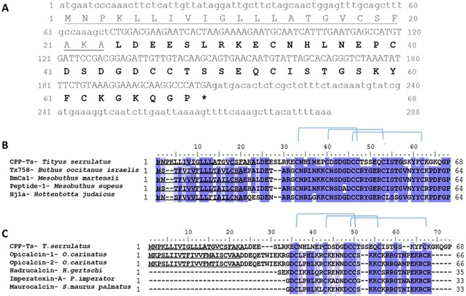
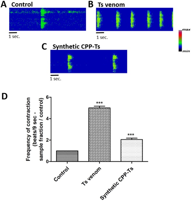
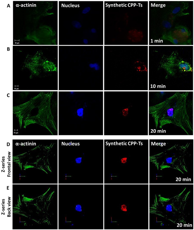
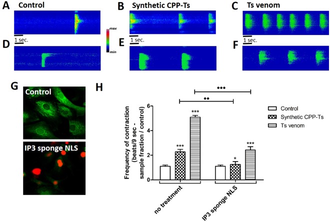
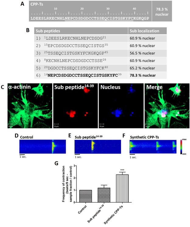
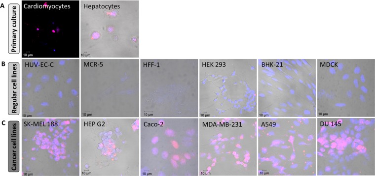
Scorpion sting envenoming impacts millions of people worldwide, with cardiac effects being one of the main causes of death on victims. Here we describe the first Ca2+ channel toxin present in Tityus serrulatus (Ts) venom, a cell penetrating peptide (CPP) named CPP-Ts. We show that CPP-Ts increases intracellular Ca2+ release through the activation of nuclear InsP3R of cardiomyocytes, thereby causing an increase in the contraction frequency of these cells. Besides proposing a novel subfamily of Ca2+ active toxins, we investigated its potential use as a drug delivery system targeting cancer cell nucleus using CPP-Ts's nuclear-targeting property. To this end, we prepared a synthetic CPP-Ts sub peptide14-39 lacking pharmacological activity which was directed to the nucleus of specific cancer cell lines. This research identifies a novel subfamily of Ca2+ active toxins and provides new insights into biotechnological applications of animal venoms.


Conflict of interest statement

The authors declare no competing interests.

Figures

Figure 1
Figure 1
Sequence of CPP-Ts from T. serrulatus venom and alignment with other scorpionic Ca2+ channel toxins. (A) Figure shows cDNA and predicted amino acid sequences of CPP-Ts (GenBank MH061344). The signal peptide sequence is underlined. Mature protein sequence is represented by the bolded amino acids and the capital nucleotides. Stop codon is represented by an asterisk. (BC) Conserved residues are marked in dark blue, similar ones in light blue, and cysteine forming disulfide bonds are connected by lines. The signal peptide sequence is underlined. (B) Alignment of T. serrulatus CPP-Ts amino acid sequence with the highest similarity sequences available in the database (60% similarity). Those are Tx758 from Buthus occitanus israelis (UniProt B8XH22), BmCa1 from Mesobuthus martensii (UniProt Q8I6X9), Peptide-1 from Mesobuthus eupeus (UniProt P86399), and Hj1a from Hottentotta judaicus (GenBank ADY39527.1). (C) CPP-Ts amino acid sequence alignment with toxins from the scorpionic calcine group (<40% similarity): Opicalcin-1 (UniProt P60252) and Opicalcin-2 (UniProt P60253) from Opistophthalmus carinatus, Hadrucalcin from Hadrurus gertschi (GenBank ACC99422.1), Imperatoxin-A from Pandinus imperator (UniProt P59868), and Maurocalcin from Scorpio maurus palmatus (UniProt P60254).
Figure 2
Figure 2
Synthetic CPP-Ts and T. serrulatus venom alter the Ca2+ transient in cardiomyocytes. Neonatal rat cardiomyocytes were used for the functional analysis of Ts venom and synthetic CPP-Ts. Ca2+ was monitored with Fluo-4/AM using confocal line-scanning microscopy (magnification x63). (AC) Global Ca2+ transient in cardiomyocytes. Cells were examined immediately after treatment with B: Ts venom (12.8 µg/ml), C: synthetic CPP-Ts (2 µg/ml). Images are pseudocolored according to the color scale. (D) Global Ca2+ transient analysis measured by the number of contractions over 9 seconds. Treatments with Ts venom and synthetic CPP-Ts significantly increased the contraction frequency in neonatal rat cardiomyocytes, when compared to control (F3,19 = 133.4, p = 6.08 e-13; tTsV = 25.12, dfTsV = 19, pTsV = 4.44 e-16; tsynth. CPP = 8.407, dfsynth. CPP = 19, psynth. CPP = 7.95 e-08). All values correspond to the mean ± S.E.M (n = 20 cells per treatment) of three independent experiments. Statistical analyses were performed using Repeated Measures ANOVA followed by paired t-tests corrected with Bonferroni procedure.
Figure 3
Figure 3
Intranuclear localization of CPP-Ts in cardiomyocytes over time. Neonatal rat cardiomyocytes were double labeled with α-actinin (green) and the nucleus marker TO-PRO-3 (blue). CPP-Ts was labeled with Alexa 555 nm (red). (AC) Confocal immunofluorescence, in single plan, shows: (A) Diffuse intracellular localization of CPP-Ts after 1 min of treatment; (B) CPP-Ts concentrated in the perinuclear region after 10 min of treatment; (C) Intranuclear localization of CPP-Ts after 20 min of treatment. It is noteworthy that CPP-Ts is driven to the nuclear region over time. (D,E) Three-dimensional images from confocal microscopy analyzed by Z-series on Volocyte program shows colocalization of CPP-Ts and the nucleus. (D) Cell frontal view with nucleus labeling adjusted for solid surface. (E) Cell back view with nuclear labeling adjusted for solid surface. It is noticeable that TO-PRO3 fluorescence completely covers Alexa 555, in all three dimensions (D,E), thus validating the intranuclear localization of CPP-Ts.
Figure 4
Figure 4
Effects of synthetic CPP-Ts and Ts venom in cardiomyocytes are reduced in cells transfected with nuclear InsP3 sponge NLS virus. Neonatal rat cardiomyocytes were transfected with IP3 sponge NLS virus and then treated with synthetic CPP-Ts or Ts venom. Experiments were conducted by monitoring of Ca2+ with Fluo-4/AM using confocal line-scanning microscopy. Global Ca2+ transient was examined immediately after treatment with CPP-Ts (2 µg/ml) or Ts venom (12.8 µg/ml) in untransfected cardiomyocytes (AC) or in cardiomyocytes transfected with nuclear IP3 sponge NLS virus (DF). Images are pseudocolored according to the color scale. (G) Representative image of both untransfected (control) and transfected cells. Intracellular Ca2+ is marked in green by Fluo-4/AM and the virus nuclear transfection is labeled in red. (H) Global Ca2+ transient analysis measured by the number of contractions over 9 seconds. Untransfected cells, treated with CPP-Ts and Ts venom (F2,19 = 616.9, p = 0; tsynth. CPP = 9.097, dfsynth. CPP = 19, psynth. CPP = 2.36 e-08***; tTsV = 40.04, dfTsV = 19, pTsV = 0***) significantly increased the contraction frequency in neonatal rat cardiomyocytes, compared to control. However, in cardiomyocytes transfected with IP3 sponge NLS virus (F2,19 = 30.82, p = 1.08 e-06; tsynth. CPP = 2.378, dfsynth. CPP = 19, psynth. CPP = 0.0281*; tTsV = 8.208, dfTsV = 19, pTsV = 1.14 e-07***), there is a significant decrease in contraction frequency compared to the untransfected group after treatment with both CPP-Ts (t = 3.454, df = 38, p = 0.0014••) or Ts venom (t = 3.42, df = 38, p = 4.44 e-16•••). This indicates that nuclear Ca2+ has an important role in the increase of global Ca2+ transient provoked by CPP-Ts and Ts venom in cardiomyocytes. All values are the mean ± S.E.M. (n = 20 cells per treatment) of three independent experiments. Statistical analyses were performed using Repeated Measures ANOVA followed by paired or unpaired t-tests corrected with Bonferroni procedure.
Figure 5
Figure 5
Biodistribution of synthetic 99mTc-CPP-Ts. (A) 99mTc-CPP-Ts (3.7 MBq) was intravenously injected in Swiss mice (n = 7, 6–8 weeks old, 24–28 g). After 10, 30, and 60 min post-injection, the radioactivity was measured in liver, spleen, kidneys, stomach, heart, lungs, blood, muscle, thyroid, intestine, brain, and pancreas. The results are expressed as the percentage of injected dose/g of tissue (%ID/g). (B) Representative scintigraphic images of mice (n = 3, 6–8 weeks old, 24–28 g) injected with 11 MBq 99mTc-CPP-Ts after 10, 30, and 60 min, showing a very high kidney uptake. (C) Comparing the tissues-to-blood ratios, there is an increasing ratio over time for heart, lungs, and liver when compared to muscle, a non-specific tissue. After 60 min, ratios reached values higher than 1.5, indicating that such organs had more than 50% of the 99mTc-CPP-Ts uptake compared to blood (organF3,72 = 282.6, p = 0; timeF2,72 = 277, p = 0; tHeart10′ = 11.90, dfHeart10′ = 6, pHeart10′ = 2.13 e-05**; tLiver10′ = 34.91, dfLiver10′ = 6, pLiver10′ = 3.6 e-08***; tLungs10′ = 15.59, dfLungs10′ = 6, pLungs10′ = 4.40 e-06***; tHeart30′ = 16.44, dfHeart30′ = 6, pHeart30′ = 3.22 e-06***; tLiver30′ = 20.73, dfLiver30′ = 6, pLiver30′ = 8.20 e-07***; tLungs30′ = 38.13, dfLungs30′ = 6, pLungs30′ = 2.17 e-08***; tHeart60′ = 29.47, dfHeart60′ = 6, pHeart60′ = 1.01 e-7***; tLiver60′ = 15.93, dfLiver60′ = 6, pLiver60′ = 3.88 e-06***; tLungs60′ = 15.47, dfLungs60′ = 6, pLungs60′ = 4.61 e-6***). Data are expressed as mean S.E.M of two independent experiments. Statistical analyses were performed using Two-way analysis of variance followed by unpaired t-tests corrected with Bonferroni procedure.
Figure 6
Figure 6
In silico prediction of the sub cellular localization and activity of sub peptide14–39 that carries the nuclear internalization property but lacks CPP-Ts biological activity. Mature CPP-Ts and six sub peptide sequences were subjected to the sub cellular localization prediction analysis using the PSORT II software. (A) CPP-Ts was accurately predicted (78.3%) to have a nuclear localization. (B) Predictions of sub cellular localization of the sub peptides varied from 56.5% to 78.3%. The sub peptide14–39 was the only one that carried the full CPP-Ts nuclear internalization properties. (C) Single plane images of neonatal rat cardiomyocytes from confocal microscopy. Merged image of cytoskeleton (green, α-actinin, 488 nm), sub peptide14–39 (red, Alexa 555, 555 nm) and nucleus (blue, TO-PRO3, 633 nm) labeling showing intranuclear localization of sub peptide14–39 after 20 min of treatment. (DG) Global Ca2+ transient analysis of cardiomyocytes stained with Fluo-4/AM using confocal line-scanning microscopy. Images are pseudocolored according to the color scale. Cells were examined immediately after treatment with sub peptide14–39 (2 µg/ml) or synthetic CPP-Ts (2 µg/ml) used as positive control. The frequency of cell contractions was altered by the treatment with synthetic CPP-Ts, but it was not significantly altered by the treatment with the sub peptide14–39 (F2,29 = 44.76, p = 1.36 e-09; tsub pep = 1.705, dfsub pep = 29, psub pep = 0.0989; tsynth. CPP = 8.572, dfsynth. CPP = 29, p synth. CPP = 1.92 e-09). All values correspond to the mean ± S.E.M (n = 20 cells per treatment) of three independent experiments. Statistical analyses were performed using Repeated Measures ANOVA followed by paired t-tests corrected with Bonferroni procedure.
Figure 7
Figure 7
Sub peptide14–39 selectivity cell internalization. The internalization property of sub peptide14–39 (20 µg/ml) was investigated in different cell lines and primary cultures. Confocal microscopy images represent the nucleus in blue (TO-PRO3, 633 nm) and sub peptide14–39 in red (Alexa 555, 555 nm). Sub peptide14–39 is able to entry in primary culture cells as cardiomyocytes and hepatocytes (A), however it is not able to entry in six normal immortalized cell lines tested: HUV-EC-C, MCR-5, HFF-1, HEK-293, BHK- 21 and MDCK (B). Interestingly, sub peptide14–39 internalizes in six cancer cell lines tested: SK-MEL-188, HEP G2, Caco-2, MDA-MB-231, A549 and DU 145 (C). The images represent two coverslips (n = 300 cells) for each cell line tested.

Similar articles

Cited by

References

    1. World health organization (WHO). Rabies and envenomings: a neglected public health issue: report of a consultative meeting. (World Health Organization, Geneva, 36 p. 2007).
    1. Chippaux JP. Emerging options for the management of scorpion stings. Drug Des Devel Ther., v. 2012;6:165–73. doi: 10.2147/DDDT.S24754. - DOI - PMC - PubMed
    1. Pucca MB, et al. Tityus serrulatus venom–A lethal cocktail. Toxicon, v. 2015;108:272–84. doi: 10.1016/j.toxicon.2015.10.015. - DOI - PubMed
    1. Ismail M. The scorpion envenoming syndrome. Toxicon, v. 1995;33:825–858. doi: 10.1016/0041-0101(95)00005-7. - DOI - PubMed
    1. Gordon D, Savarin P, Gurevitz M, Zinn-Justin S. Functional anatomy of scorpion toxins affecting sodium channels. J Toxicol Toxin Rev., v. 1998;17:131–159. doi: 10.3109/15569549809009247. - DOI

Publication types