. 2018 Oct 3;9(1):4049.
doi: 10.1038/s41467-018-06183-4.

Bi-directional cell-pericellular matrix interactions direct stem cell fate

Affiliations

Bi-directional cell-pericellular matrix interactions direct stem cell fate

Silvia A Ferreira et al. Nat Commun. .

Erratum in

  • Author Correction: Bi-directional cell-pericellular matrix interactions direct stem cell fate.
    Ferreira SA, Motwani MS, Faull PA, Seymour AJ, Yu TTL, Enayati M, Taheem DK, Salzlechner C, Haghighi T, Kania EM, Oommen OP, Ahmed T, Loaiza S, Parzych K, Dazzi F, Varghese OP, Festy F, Grigoriadis AE, Auner HW, Snijders AP, Bozec L, Gentleman E. Ferreira SA, et al. Nat Commun. 2018 Nov 14;9(1):4851. doi: 10.1038/s41467-018-07398-1. Nat Commun. 2018. PMID: 30429483 Free PMC article.
  • Author Correction: Bi-directional cell-pericellular matrix interactions direct stem cell fate.
    Ferreira SA, Motwani MS, Faull PA, Seymour AJ, Yu TTL, Enayati M, Taheem DK, Salzlechner C, Haghighi T, Kania EM, Oommen OP, Ahmed T, Loaiza S, Parzych K, Dazzi F, Varghese OP, Festy F, Grigoriadis AE, Auner HW, Snijders AP, Bozec L, Gentleman E. Ferreira SA, et al. Nat Commun. 2018 Dec 18;9(1):5419. doi: 10.1038/s41467-018-07843-1. Nat Commun. 2018. PMID: 30560926 Free PMC article.

Abstract

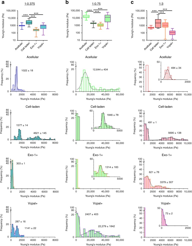
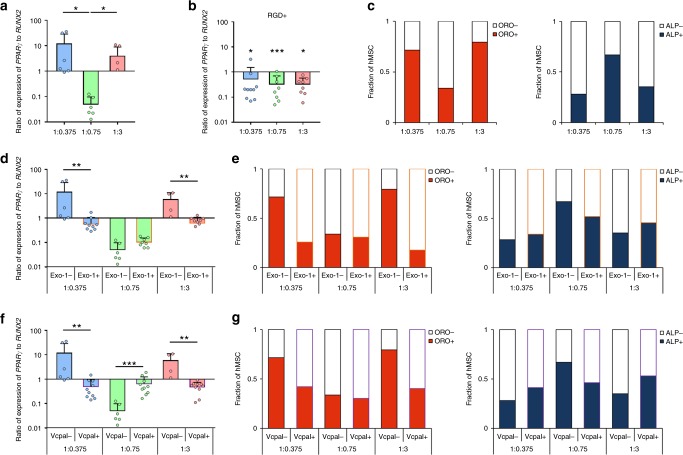
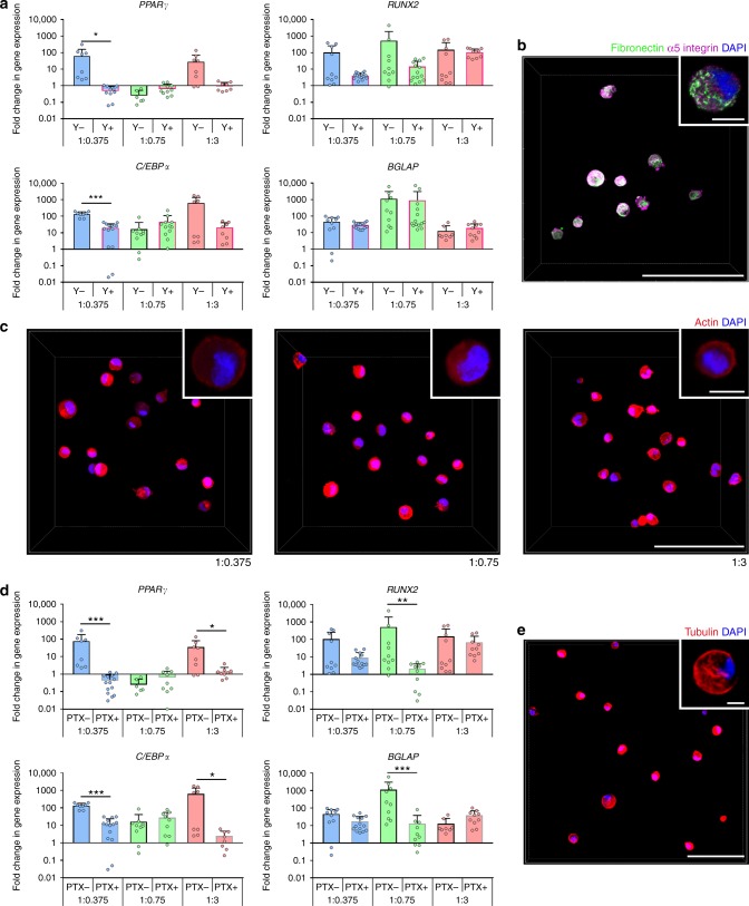
Modifiable hydrogels have revealed tremendous insight into how physical characteristics of cells' 3D environment drive stem cell lineage specification. However, in native tissues, cells do not passively receive signals from their niche. Instead they actively probe and modify their pericellular space to suit their needs, yet the dynamics of cells' reciprocal interactions with their pericellular environment when encapsulated within hydrogels remains relatively unexplored. Here, we show that human bone marrow stromal cells (hMSC) encapsulated within hyaluronic acid-based hydrogels modify their surroundings by synthesizing, secreting and arranging proteins pericellularly or by degrading the hydrogel. hMSC's interactions with this local environment have a role in regulating hMSC fate, with a secreted proteinaceous pericellular matrix associated with adipogenesis, and degradation with osteogenesis. Our observations suggest that hMSC participate in a bi-directional interplay between the properties of their 3D milieu and their own secreted pericellular matrix, and that this combination of interactions drives fate.


Conflict of interest statement

The authors declare no competing interests.

Figures

Fig. 1
Fig. 1
hMSC within S-HA-PEGDA hydrogels synthesize and secrete proteins pericellularly. a Reaction scheme for hydrogel formation. Thiol-modified hyaluronic acid (S-HA) cross-links with poly(ethylene glycol) diacrylate (PEGDA) to form a hydrogel via a Michael addition. b Viability of hMSC encapsulated in 1:0.75 hydrogels treated with an anti-CD44 (CD44+) antibody or an isotype control (CD44−) for 24 h and normalized to vehicle controls (n ≥ 3). c Viability of hMSC encapsulated in 1:0.75 hydrogels treated for 24 h with RGD sequence-containing peptides (RGD+) or scrambled peptides (RGD−) and normalized to vehicle controls (n ≥ 3). In b and c plots show means ± s.d. d Representative micrographs showing fluorescence labeling of the methionine analogue l-homopropargylglycine (HPG, green) in hMSC-laden 1:0.375, 1:0.75, and 1:3 hydrogels after 72 h in culture. The cell membrane, as determined by differential interference contrast (DIC) imaging, is outlined in white. Scale bar = 100 µm, inset = 10 µm. Plots show mean intensity of the fluorescence signal (±s.e.m.) as a function of distance from the cell membrane in 1:0.375 (blue dashes), 1:0.75 (green dashes), and 1:3 (red dashes) hydrogels and were generated from 40 profile plots collected from 30 cells per condition. e Fraction of SILAC heavy-labeled proteins in decellularized 1:0.375 (blue circles), 1:0.75 (green circles), and 1:3 (red circles) hydrogels cultured for 72 h. Scatterplots show two technical replicates (H/L count ≥ 3) for each hydrogel composition. Gene names for ECM proteins showing high levels (>40%) of SILAC incorporation are highlighted in each panel
Fig. 2
Fig. 2
Hydrogel stiffness is modified by encapsulated hMSC. Young’s modulus (E, Pa) of acellular and cell-laden a 1:0.375, b 1:0.75, and c 1:3 S-HA-PEGDA hydrogels evaluated by atomic force microscopy. Box plots (top row) showing 1st/3rd quartiles (bounds of box), high/low values (whiskers), and median (central line) E. A Mann–Whitney test (two-tailed) was used to assess statistical differences (***p < 0.001). Histograms show distributions of E on acellular and hMSC-laden hydrogels cultured under standard conditions or treated with 75 μM Exo-1 or 100 μM Vcpal for 72 h. Histograms are overlaid with fitted normal distributions, with centre ± standard error. Insets show distributions and fitted normal distributions in designated areas of the datasets. In all compositions, the presence of hMSC significantly affected the distribution of E (see Supplementary Table 2 for n values and statistical analyses of distributions of E)
Fig. 3
Fig. 3
hMSC’s modifications to their pericellular environment directs fate. a Ratio of expression of PPARγ to RUNX2 in hMSC encapsulated in hydrogels for 72 h and normalized to undifferentiated hMSC controls (mean + s.d.). Ratios were significantly higher in 1:0.375 and 1:3 (*p < 0.05) compared to 1:0.75 hydrogels (n ≥ 4). b Ratio of expression of PPARγ to RUNX2 in hMSC after treatment with RGD sequence-containing peptides (RGD+) for 72 h (mean + s.d.). Ratios were not significantly different in hydrogels of different compositions (n ≥ 8), but for each composition, expression was significantly different than RGD− controls (1:0.375 and 1:3, *p < 0.05; 1:0.75, ***p < 0.001). c Fraction of hMSC that stained positively for Oil Red O (ORO+) or alkaline phosphatase (ALP+), as indicators of adipogenesis and osteogenesis, respectively, after 14 days in culture in a bi-potential osteogenic/adipogenic medium (n = 3300–1200 cells). d Ratio of expression of PPARγ to RUNX2 in hMSC after treatment with 75 μM Exo-1 or f 100 μM Vcpal for 72 h (mean + s.d.). Fraction of hMSC that stained positively for ORO and ALP after treatment with either e 75 μM Exo-1 or g 100 μM Vcpal for 14 days (n = 3300–1200 cells). In a a Kruskal–Wallis followed by Dunn’s multiple comparison test was used to detect statistical significance. In bd, and f a Mann–Whitney test (two-tailed) was used to compare treatment (RGD+, Exo-1+, Vcpal+) to control conditions within each hydrogel composition. In ce, and g a Fisher’s exact test (two-sided) was used to compare treatment (Exo-1+, Vcpal+) to control conditions within each hydrogel composition (Supplementary Table 3). For representative images of ORO and ALP staining, see Supplementary Fig. 13
Fig. 4
Fig. 4
Y-27632 and paclitaxel impact osteogenic/adipogenic gene expression. a Gene expression analyses for markers of adipogenesis (PPARγ and C/EPBα) and osteogenesis (RUNX2 and BGLAP) in hMSC cultured for 72 h with basal culture medium (−Y) or treated with 10 μM Y-27632 (+Y, n ≥ 8). Expression levels (mean + s.d.) are shown as fold change normalized to expression in undifferentiated hMSC (set to 1). b Representative micrograph of hMSC (5 × 106 cells mL−1) encapsulated in a 1:0.75 hydrogel after 28 days showing fibronectin, α5 integrin, and DAPI. Fibronectin co-localized with punctate α5 integrin by Manders’ coefficients: Manders’ tM1 = 0.83 ± 0.18 and Manders’ tM2 = 0.84 ± 0.10 (n = 4, 25 cells). c Representative micrographs of hMSC within hydrogels after 72 h and stained with Phalloidin-TRITC (actin) and DAPI. d Gene expression analyses for markers of adipogenesis (PPARγ and C/EPBα) and osteogenesis (RUNX2 and BGLAP) in hMSC cultured for 72 h with basal culture medium (−PTX) or treated with 50 nM paclitaxel (+PTX, n ≥ 8). Expression levels (mean + s.d.) are shown as fold change normalized to expression in undifferentiated hMSC (set to 1). e Representative micrographs of hMSC (5 × 106 cells mL−1) within a 1:0.75 hydrogel for 72 h and stained for tubulin and DAPI. Scale bars in b, c, and e are 100 µm, and in insets = 10 µm. In a and d a Mann–Whitney test (two-tailed) was used to detect statistical significance, *p < 0.05, **p < 0.01, ***p < 0.001

Similar articles

Cited by

References

    1. Walters NJ, Gentleman E. Evolving insights in cell-matrix interactions: elucidating how non-soluble properties of the extracellular niche direct stem cell fate. Acta Biomater. 2015;11:3–16. doi: 10.1016/j.actbio.2014.09.038. - DOI - PMC - PubMed
    1. Kloxin AM, Kasko AM, Salinas CN, Anseth KS. Photodegradable hydrogels for dynamic tuning of physical and chemical properties. Science. 2009;324:59–63. doi: 10.1126/science.1169494. - DOI - PMC - PubMed
    1. Lutolf MP, Hubbell JA. Synthetic biomaterials as instructive extracellular microenvironments for morphogenesis in tissue engineering. Nat. Biotechnol. 2005;23:47–55. doi: 10.1038/nbt1055. - DOI - PubMed
    1. Huebsch N, et al. Harnessing traction-mediated manipulation of the cell/matrix interface to control stem-cell fate. Nat. Mater. 2010;9:518–526. doi: 10.1038/nmat2732. - DOI - PMC - PubMed
    1. Khetan S, et al. Degradation-mediated cellular traction directs stem cell fate in covalently crosslinked three-dimensional hydrogels. Nat. Mater. 2013;12:458–465. doi: 10.1038/nmat3586. - DOI - PMC - PubMed

Publication types

MeSH terms