. 2018 Oct 3;8(1):14736.
doi: 10.1038/s41598-018-32938-6.

Profiling inflammatory markers in patients with pneumonia on intensive care

Affiliations

Profiling inflammatory markers in patients with pneumonia on intensive care

David B Antcliffe et al. Sci Rep. .

Abstract

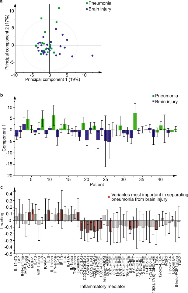
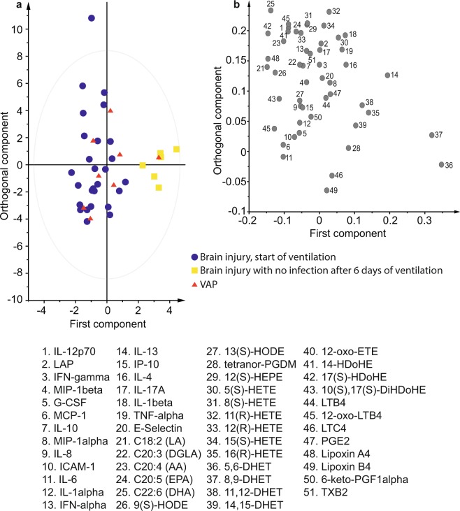
Clinical investigations lack predictive value when diagnosing pneumonia, especially when patients are ventilated and develop ventilator associated pneumonia (VAP). New tools to aid diagnosis are important to improve outcomes. This pilot study examines the potential for a panel of inflammatory mediators to aid in the diagnosis. Forty-four ventilated patients, 17 with pneumonia and 27 with brain injuries, eight of whom developed VAP, were recruited. 51 inflammatory mediators, including cytokines and oxylipins, were measured in patients' serum using flow cytometry and mass spectrometry. The mediators could separate patients admitted to ICU with pneumonia compared to brain injury with an area under the receiver operating characteristic curve (AUROC) 0.75 (0.61-0.90). Changes in inflammatory mediators were similar in both groups over the course of ICU stay with 5,6-dihydroxyeicosatrienoic and 8,9-dihydroxyeicosatrienoic acids increasing over time and interleukin-6 decreasing. However, brain injured patients who developed VAP maintained inflammatory profiles similar to those at admission. A multivariate model containing 5,6-dihydroxyeicosatrienoic acid, 8,9-dihydroxyeicosatrienoic acid, intercellular adhesion molecule-1, interleukin-6, and interleukin-8, could differentiate patients with VAP from brain injured patients without infection (AUROC 0.94 (0.80-1.00)). The use of a selected group of markers showed promise to aid the diagnosis of VAP especially when combined with clinical data.


Conflict of interest statement

The authors declare no competing interests.

Figures

Figure 1
Figure 1
Multivariate analysis using all inflammatory mediators comparing samples taken at admission from patients admitted with pneumonia to those admitted with brain injuries. (a) Principal component analysis scores plot comparing principal components 1 and 2 showing pneumonia (green squares) and those admitted with brain injuries (blue circles). (b) Orthogonal partial least squares discriminant analysis (OPLS-DA) model with one component (R2Y 0.38 Q2 0.22, p = 0.007) with pneumonia represented by green bars and brain injury by blue bars. Pneumonia samples project in a positive direction along the first component, y-axis, and brain injury samples in the negative direction. (c) Loadings plot for the model shown in b, inflammatory molecules at the top of the plot dominate in the pneumonia group and those towards the bottom in the brain injury group. Those marked in red had the greatest influence on the model. Error bars in b and c represent jack-knifed 95% standard error of the scores and loadings respectively.
Figure 2
Figure 2
Change in concentration of 5,6-dihydroxyeicosatrienoic (5,6-DHET) (a) and Intercellular Adhesion Molecule-1 (ICAM-1) (b) over time on Intensive Care for patients with pneumonia (dark line), brain injury without infection (pale line) and brain injury who develop ventilator associated pneumonia (VAP) (dashed line). VAP represents the time that VAP was diagnosed which was not always the final time point. VAP is compared to the day 6 time point in those without infection as this was the timing of diagnosis in 50% of cases. Data are displayed as median with interquartile range.
Figure 3
Figure 3
Orthogonal partial least squares discriminant analysis (OPLS-DA) comparing admission samples in patients with brain injuries to those taken after 6 days of ventilation. (a) OPLS-DA scores plot comparing samples taken from brain injured patients at the start of ventilation (blue circles) and after a further 6 days of ventilation for those without infection (yellow squares). Samples taken from patients with ventilator associated pneumonia have been predicted by the model to lie mainly amongst the first time point samples (red triangles). (b) Corresponding loadings plot for the OPLS-DA model showing the mediators that account for most of the difference between the groups. Mediators to the right of the x-axis dominate in the final time point of those without infection where as those to the far left dominate at the start of ventilation. 5,6-dihydroxyeicosatrienoic (5,6-DHET) and 8,9-dihydroxyeicosatrienoic (8,9-DHET) are the most influential oxylipins in the final time point samples.

Similar articles

Cited by

References

    1. Martin-Loeches I, et al. Incidence and prognosis of ventilator-associated tracheobronchitis (TAVeM): a multicentre, prospective, observational study. Lancet Respir. Med. 2015;3:859–868. doi: 10.1016/S2213-2600(15)00326-4. - DOI - PubMed
    1. Chastre J, Fagon JY. Ventilator-associated pneumonia. Am. J. Respir. Crit. Care Med. 2002;165:867–903. doi: 10.1164/ajrccm.165.7.2105078. - DOI - PubMed
    1. Kollef MH. Ventilator-associated pneumonia. A multivariate analysis. JAMA. 1993;270:1965–1970. doi: 10.1001/jama.1993.03510160083034. - DOI - PubMed
    1. Rello J, et al. Epidemiology and outcomes of ventilator-associated pneumonia in a large US database. Chest. 2002;122:2115–2121. doi: 10.1378/chest.122.6.2115. - DOI - PubMed
    1. Fagon JY, et al. Evaluation of clinical judgment in the identification and treatment of nosocomial pneumonia in ventilated patients. Chest. 1993;103:547–553. doi: 10.1378/chest.103.2.547. - DOI - PubMed

Publication types