. 2018 Oct 3;8(1):14749.
doi: 10.1038/s41598-018-33083-w.

Graphitic Carbon Electrodes on Flexible Substrate for Neural Applications Entirely Fabricated Using Infrared Nanosecond Laser Technology

Affiliations

Graphitic Carbon Electrodes on Flexible Substrate for Neural Applications Entirely Fabricated Using Infrared Nanosecond Laser Technology

Maria Vomero et al. Sci Rep. .

Abstract

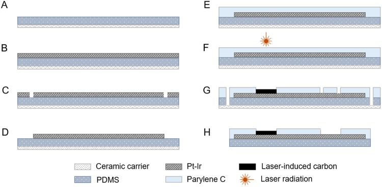
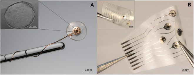
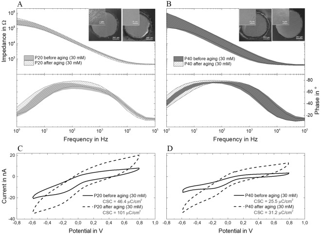
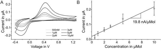
Neural interfaces for neuroscientific research are nowadays mainly manufactured using standard microsystems engineering technologies which are incompatible with the integration of carbon as electrode material. In this work, we investigate a new method to fabricate graphitic carbon electrode arrays on flexible substrates. The devices were manufactured using infrared nanosecond laser technology for both patterning all components and carbonizing the electrode sites. Two laser pulse repetition frequencies were used for carbonization with the aim of finding the optimum. Prototypes of the devices were evaluated in vitro in 30 mM hydrogen peroxide to mimic the post-surgery oxidative environment. The electrodes were subjected to 10 million biphasic pulses (39.5 μC/cm2) to measure their stability under electrical stress. Their biosensing capabilities were evaluated in different concentrations of dopamine in phosphate buffered saline solution. Raman spectroscopy and x-ray photoelectron spectroscopy analysis show that the atomic percentage of graphitic carbon in the manufactured electrodes reaches the remarkable value of 75%. Results prove that the infrared nanosecond laser yields activated graphite electrodes that are conductive, non-cytotoxic and electrochemically inert. Their comprehensive assessment indicates that our laser-induced carbon electrodes are suitable for future transfer into in vivo studies, including neural recordings, stimulation and neurotransmitters detection.


Conflict of interest statement

The authors declare no competing interests.

Figures

Figure 1
Figure 1
Schematic of the fabrication process of the electrode arrays. (A) Spin-coating of silicone rubber onto a ceramic carrier. (B) Lamination of the metal layer. (C) Structuring of the metal. (D) Removal of the metal excess. (E) Parylene C coating. (F) Laser pyrolysis of the active site and structuring of the electrodes (700 µm in diameter). (G) Opening of the metal pads. (H) Device releasing.
Figure 2
Figure 2
Pictures of (A) a single-channel device - with SEM (scanning electron microscopy) picture of the carbon electrode in the inset - and of (B) an ultra-flexible 9-electrode array (curled in the inset). Both were manufactured with the method described above.
Figure 3
Figure 3
EIS measurements (magnitude and phase) of four electrodes per group, P20 (A) and P40 (B), before and after immersion in 30 mM H2O2 for one week at 37 °C. Shade regions consider the average and standard deviation of the obtained data points. Cyclic voltammograms of a (C) P20 and (D) P40 electrode (C) before and after immersion in 30 mM H2O2 for one week at 37 °C.
Figure 4
Figure 4
EIS measurements (magnitude and phase) of a representative electrode of each group - P20 (A) and P40 (B) - before the electrical stimulation test and after 1, 5 and 10 million biphasic pulses. Cyclic voltammograms of electrodes P20 (C) and P40 (D), before the electrical stimulation test and after 1, 5 and 10 million biphasic pulses.
Figure 5
Figure 5
SEM images of a P40-type laser-induced carbon electrode after electrical stimulation (10 million biphasic current pulses). No delamination is observed in the overall picture (A) nor in the detail (B) taken at 1000x magnification.
Figure 6
Figure 6
(A) Wide-scan or survey spectrum of a P20 laser-induced carbon electrode, showing all elements present; (B) XPS spectra of C 1s energy level for the same P20 sample. (C) Survey spectrum of a P40 laser-induced carbon electrode and (D) XPS spectra of C 1s energy level for the same P40 sample.
Figure 7
Figure 7
Raman spectroscopy of pristine electrodes P20 (top) and P40 (bottom). Three measurements were taken for each sample (in three random locations).
Figure 8
Figure 8
Quantitative analysis of the viability of neural cells cultured for 24 h in medium previously incubated with samples of laser-induced carbon P20 and P40 as well as normal cell medium (negative control). In the positive control, the cells were intentionally exposed to a toxic medium.
Figure 9
Figure 9
(A) Background-subtracted voltammograms of a representative P20-type carbon electrode when subjected to different dopamine concentrations (500 nM, 1 µM, 2 µM, 3 µM, 5 µM and 10 µM) in phosphate buffered saline solution and (B) calibration curve (grey line) obtained by plotting the average peak current (with standard deviation) over the concentrations of dopamine detected in vitro using three P20-type carbon electrodes (200 µm in diameter).

References

    1. Polikov VS, Tresco PA, Reichert WM. Response of brain tissue to chronically implanted neural electrodes. J. Neurosc. Meth. 2005;148:1–18. doi: 10.1016/j.jneumeth.2005.08.015. - DOI - PubMed
    1. Biran R, Martin DC, Tresco PA. Neuronal cell loss accompanies the brain tissue response to chronically implanted silicon microelectrode arrays. Exp Neurol. 2005;195:115–26. doi: 10.1016/j.expneurol.2005.04.020. - DOI - PubMed
    1. Williams JC, Hippensteel JA, Dilgen J, Shain W, Kipke DR. Complex impedance spectroscopy for monitoring tissue responses to inserted neural implants. J. Neu. Eng. 2007;4:410–423. doi: 10.1088/1741-2560/4/4/007. - DOI - PubMed
    1. Biran R, Martin DC, Tresco PA. The brain tissue response to implanted silicon microelectrode arrays is increased when the device is tethered to the skull. J. Biom. Mat. Res. 2007;82A:169–178. doi: 10.1002/jbm.a.31138. - DOI - PubMed
    1. Turner JN, et al. H. Cerebral astrocyte response to micromachined silicon implants. Exp. Neurol. 1999;156:33–49. doi: 10.1006/exnr.1998.6983. - DOI - PubMed

Publication types