. 2018 Oct 3;8(1):14728.
doi: 10.1038/s41598-018-33016-7.

Activity of a novel antimicrobial peptide against Pseudomonas aeruginosa biofilms

Affiliations

Activity of a novel antimicrobial peptide against Pseudomonas aeruginosa biofilms

Trevor Beaudoin et al. Sci Rep. .

Abstract

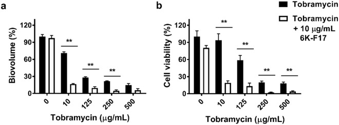
With the increasing recognition of biofilms in human disease, the development of novel antimicrobial therapies is of critical importance. For example, in patients with cystic fibrosis (CF), the acquisition of host-adapted, chronic Pseudomonas aeruginosa infection is associated with a decline in lung function and increased mortality. Our objective was to test the in vitro efficacy of a membrane-active antimicrobial peptide we designed, termed 6K-F17 (sequence: KKKKKK-AAFAAWAAFAA-NH2), against multidrug resistant P. aeruginosa biofilms. This peptide displays high antimicrobial activity against a range of pathogenic bacteria, yet is non-hemolytic to human erythrocytes and non-toxic to human bronchial epithelial cells. In the present work, P. aeruginosa strain PAO1, and four multidrug resistant (MDR) isolates from chronically infected CF individuals, were grown as 48-hour biofilms in a static biofilm slide chamber model. These biofilms were then exposed to varying concentrations of 6K-F17 alone, or in the presence of tobramycin, prior to confocal imaging. Biofilm biovolume and viability were assessed. 6K-F17 was able to kill biofilms - even in the presence of sputum - and greatly reduce biofilm biovolume in PAO1 and MDR isolates. Strikingly, when used in conjunction with tobramycin, low doses of 6K-F17 significantly potentiated tobramycin killing, leading to biofilm destruction.


Conflict of interest statement

The authors declare no competing interests.

Figures

Figure 1
Figure 1
Properties of designed cationic antimicrobial peptide 6K-F17. 6K-F17 toxicity against cystic fibrosis bronchial epithelial (CFBE) cells with the F508del mutation. CFBE F508del cells were incubated overnight with increasing concentrations of 6K-F17, and toxicity was assessed by the fluorescence of Calcein-AM. The percentage of live cells was normalized to the fluorescence of cells treated with cell culture media alone (0.0 µg/mL peptide). 1% SDS detergent, and 20 µg/mL puromycin (Puro), were used as positive controls for cell death. A visual representation of the assay is shown above the graph, where live cells are depicted in pink, dead cells in blue. Values represent the average from N = 3 biological replicates; error is reported as standard error of the mean.
Figure 2
Figure 2
Cationic peptide 6K-F17 kills P. aeruginosa biofilms. (a) Representative images of 30-hour slide chamber P. aeruginosa PAO1 biofilms were grown with increasing concentrations of 6K-F17 for 16 hours prior to confocal imaging and analysis of biovolume. (b) Total biofilm biovolume expressed as percentage of untreated controls. The mean of n = 12 images from N = 4 biological experiments is plotted. Comparison of means between conditions was performed using a Kruskal-Wallis test with a Dunn's multiple comparison post-test. (c) ATP cell viability assay of PAO1 biofilms exposed to increasing concentrations of 6K-F17. PAO1 was grown in 96-well microtiter plates and ATP assays were performed as described in the methods. The mean of n = 4 experiments is plotted. Comparison of means between conditions was performed using a Mann-Whitney U test, **p < 0.01. Error is reported as standard error of the mean.
Figure 3
Figure 3
Cationic peptide 6K-F17 potentiates tobramycin activity against P. aeruginosa biofilms. 30-hour slide chamber P. aeruginosa PAO1 biofilms were grown with tobramycin alone in increasing concentrations, or with a fixed concentration (10 µg/mL) of 6K-F17 + increasing concentrations of tobramycin for 16 hours prior to confocal imaging and analysis of biovolume. (a) Total biofilm biovolume expressed as percentage of untreated controls. The mean of n = 12 images from N = 4 biological experiments is plotted. Comparison of means between conditions was performed using a Kruskal-Wallis test with a Dunn’s multiple comparison post-test. (b) ATP cell viability assay of PAO1 biofilms exposed to increasing concentrations of tobramycin alone fixed concentration (10 µg/mL) of 6K-F17 + increasing concentrations of tobramycin for 16 hours. PAO1 was grown in 96-well microtiter plates and ATP assays were performed as described in the methods. The mean of n = 4 experiments is plotted. Comparison of means between conditions was performed using a Mann-Whitney U test, **p < 0.01. Error is reported as standard error of the mean.
Figure 4
Figure 4
Time course for tobramycin biofilm kill compared to 6K-F17 + tobramycin. (a) Representative images of PAO1 biofilms exposed to 6K-F17. PAO1 was grown on slide chamber in LB media for 30 hours prior to exposure with 1000 μg/mL of tobramycin alone or 10 µg/mL of 6K-F17 + 1000 μg/mL tobramycin for increasing amounts of time. Following this, biofilms were stained with a live (green cells)/dead (red cells) cell viability kit for 1 hour prior to confocal imaging. 3D images were constructed using Volocity software. (b) Total biovolume expressed as a percentage of the untreated control condition. (c) Total biovolume that is dead/total biovolume. Note that % death decreases as total biovolume decreases. The mean of n = 12 images from N = 4 biological replicates is plotted. Comparison of means compared to untreated control was performed using a Kruskal-Wallis test with a Dunn’s multiple comparison post-test, *p < 0.05, **p < 0.01, ***p < 0.001. Error is reported as standard error of the mean.
Figure 5
Figure 5
6K-F17 potentiates tobramycin activity in the presence of CF sputum. PAO1 was grown on a slide chamber in LB media alone or media with pooled sputum supernatant (10% v/v) for 48 hours prior to exposure with 100 μg/mL of tobramycin alone, or 10 μg/mL of 6K-F17 alone, or 100 μg/mL tobramycin + 10 μg/mL of 6K-F17 for 3 hours. Following this, biofilms were stained with a live/dead cell viability kit for 1 hour prior to confocal imaging. 3D images were constructed using Volocity software. (a) Total biovolume expressed as a percentage of the untreated control condition from the experiment. (b) Total biovolume that is dead/total biovolume. The mean n = 12 images from N = 4 biological replicates is shown. Comparison of means to the untreated condition was performed using a Kruskal-Wallis test with a Dunn’s multiple comparison post-test, *p < 0.05, **p < 0.01. Error is reported as standard error of the mean.
Figure 6
Figure 6
Cationic peptide 6K-F17 kills of multidrug resistant clinical isolates of P. aeruginosa biofilms. (ad) Four multidrug resistant clinical P. aeruginosa isolates from chronically infected CF patients, were grown on a slide chamber in LB media for 30 hours prior to exposure with 6K-F17 for 16 hours prior to staining and confocal imaging. Left panels illustrate the biovolume as a percent of the untreated control. The mean of n = 12 images from N = 4 biological experiments is plotted and compared between conditions using a Kruskal-Wallis test with a Dunn’s multiple comparison post-test. Right panels illustrate the ATP bacterial viability assay (mean of n = 4 experiments). Comparison of means between the two conditions was done using a Mann-Whitney U test, **p < 0.01. Error is reported as standard error of the mean.
Figure 7
Figure 7
Cationic peptide 6K-F17 potentiates tobramycin kill of multidrug resistant clinical isolates of P. aeruginosa biofilms. (a–d) Four multidrug resistant clinical P. aeruginosa isolates from chronically infected CF patients, were grown on a slide chamber in LB media for 30 hours prior to exposure with tobramycin alone in increasing concentrations, or with a fixed concentration (10 µg/mL) of 6K-F17 + increasing concentrations of tobramycin for 16 hours prior to staining and confocal imaging. Left panels illustrate the biovolume as a percent of the untreated control. The mean of n = 12 images from N = 4 biological experiments is plotted and compared between conditions using a Kruskal-Wallis test with a Dunn’s multiple comparison post-test. Right panels illustrate the ATP bacterial viability assay (mean of n = 4 experiments). Comparison of means between the two conditions was done using a Mann-Whitney U test, **p < 0.01. Error is reported as standard error of the mean.

Similar articles

Cited by

References

    1. Choo-Kang LR, Zeitlin PL. Type I, II, III, IV, and V cystic fibrosis transmembrane conductance regulator defects and opportunities for therapy. Curr. Opin. Pulm. Med. 2000;6:521–529. doi: 10.1097/00063198-200011000-00011. - DOI - PubMed
    1. Ratjen F, Döring G. Cystic fibrosis. Lancet. 2003;361:681–689. doi: 10.1016/S0140-6736(03)12567-6. - DOI - PubMed
    1. Cohen TS, Prince A. Cystic fibrosis: a mucosal immunodeficiency syndrome. Nat. Med. 2012;18:509–519. doi: 10.1038/nm.2715. - DOI - PMC - PubMed
    1. Cystic Fibrosis Canada. Canadian Patient Data Registry Report. (Toronto, Canada, 2017).
    1. Bhagirath AY, et al. Cystic fibrosis lung environment and Pseudomonas aeruginosa infection. BMC Pulm. Med. 2016;16:174. doi: 10.1186/s12890-016-0339-5. - DOI - PMC - PubMed

Publication types

MeSH terms