. 2019 Apr;24(4):588-600.
doi: 10.1038/s41380-018-0243-x. Epub 2018 Oct 3.

Adolescent exposure to Δ9-tetrahydrocannabinol alters the transcriptional trajectory and dendritic architecture of prefrontal pyramidal neurons

Affiliations

Adolescent exposure to Δ9-tetrahydrocannabinol alters the transcriptional trajectory and dendritic architecture of prefrontal pyramidal neurons

Michael L Miller et al. Mol Psychiatry. 2019 Apr.

Abstract

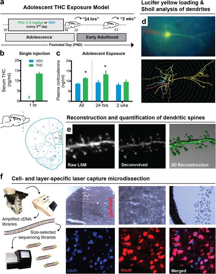
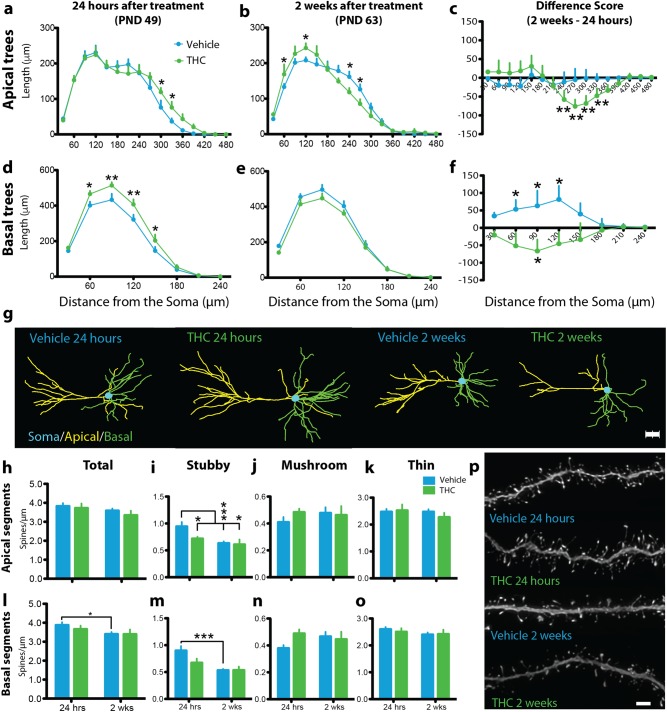
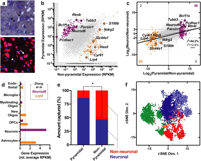
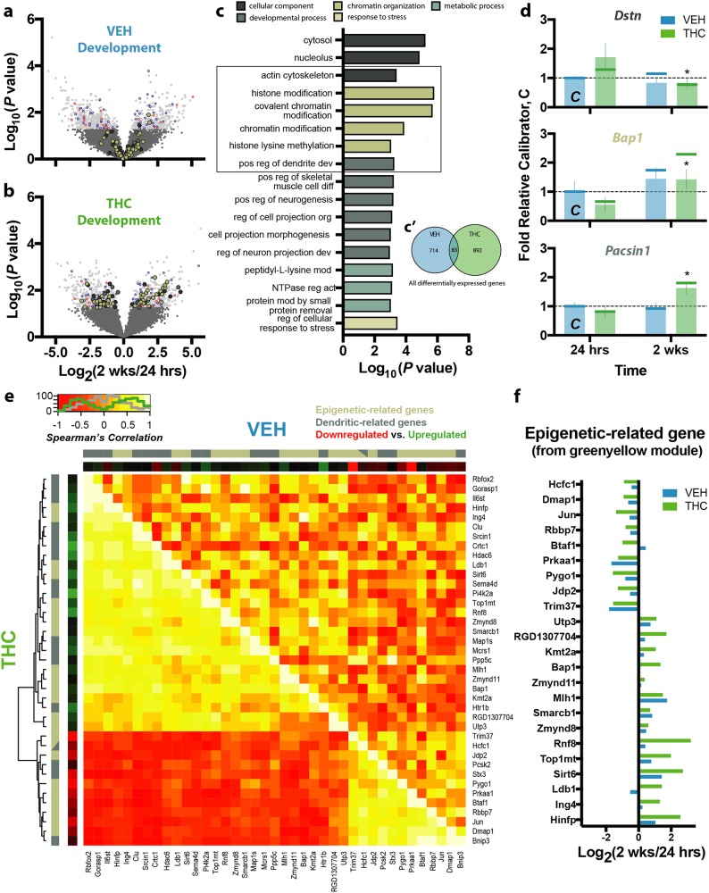
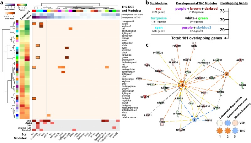
Neuronal circuits within the prefrontal cortex (PFC) mediate higher cognitive functions and emotional regulation that are disrupted in psychiatric disorders. The PFC undergoes significant maturation during adolescence, a period when cannabis use in humans has been linked to subsequent vulnerability to psychiatric disorders such as addiction and schizophrenia. Here, we investigated in a rat model the effects of adolescent exposure to Δ9-tetrahydrocannabinol (THC), a psychoactive component of cannabis, on the morphological architecture and transcriptional profile of layer III pyramidal neurons-using cell type- and layer-specific high-resolution microscopy, laser capture microdissection and next-generation RNA-sequencing. The results confirmed known normal expansions in basal dendritic arborization and dendritic spine pruning during the transition from late adolescence to early adulthood that were accompanied by differential expression of gene networks associated with neurodevelopment in control animals. In contrast, THC exposure disrupted the normal developmental process by inducing premature pruning of dendritic spines and allostatic atrophy of dendritic arborization in early adulthood. Surprisingly, there was minimal overlap of the developmental transcriptomes between THC- and vehicle-exposed rats. THC altered functional gene networks related to cell morphogenesis, dendritic development, and cytoskeleton organization. Marked developmental network disturbances were evident for epigenetic regulators with enhanced co-expression of chromatin- and dendrite-related genes in THC-treated animals. Dysregulated PFC co-expression networks common to both the THC-treated animals and patients with schizophrenia were enriched for cytoskeletal and neurite development. Overall, adolescent THC exposure altered the morphological and transcriptional trajectory of PFC pyramidal neurons, which could enhance vulnerability to psychiatric disorders.


Conflict of interest statement

The authors declare that they have no conflict of interest.

Figures

Fig. 1
Fig. 1
Model of adolescent THC exposure and approaches for layer- and cell-specific morphological and transcriptional profiling of prelimbic (PrL) pyramidal neurons. a Schematic timeline of THC exposure during adolescence and developmental time points studied. b Acute administration of 1.5 mg/kg THC increased circulating serum THC concentration 1 h after injection. c Adolescent exposure to THC elevated corticosterone levels, most notably 24 h after the last administration that may reflect a component of acute withdrawal. df Multidisciplinary approach to study the developmental effects of THC on pyramidal neuron morphology and gene expression in layer III of the rodent PrL. Dendritic branching d and spine density e were studied by filling pyramidal neurons with Lucifer yellow. (f) Pyramidal neurons from layer III were microdissected from Nissl-stained sections and sequenced for downstream transcriptome-wide profiling
Fig. 2
Fig. 2
Adolescent THC exposure altered the dendritic arborization, spine density, and developmental trajectory of layer III prelimbic (PrL) pyramidal neurons. a Adolescent THC significantly increased distal apical dendritic arborization in early adolescence (24 h after drug treatment). b In early adulthood (2 weeks after drug treatment), a significant allostatic reversal resulting in atrophy was observed in distal apical trees accompanied by the emergence of significantly increased proximal branching. c Between time points, vehicle-treated animals (VEH) exhibited no difference in apical arbor complexity while THC-treated animals (THC) exhibited a significant decrease in distal apical branching complexity. d Adolescent THC exposure significantly increased basal dendritic arborization 24 h after drug treatment. e Two weeks after drug treatment, a non-significant allostatic reversal was observed in basal trees. f Vehicle-treated animals exhibited significantly increased basal arbor complexity between time points, whereas THC-treated animals exhibited significantly decreased basal apical branching complexity. g Representative dendrograms of traced PrL pyramidal neurons. h No significant differences were observed in total apical dendritic spine density. i Vehicle-treated subjects exhibited significant pruning of stubby apical dendritic spines between time points, whereas THC-treated subjects exhibited premature pruning. j No significant differences in apical mushroom spine density were observed. k No significant differences in apical thin spine density were observed. l Vehicle-treated subjects exhibited significant pruning of total basal dendritic spines between time points, whereas THC-treated subjects exhibited premature pruning. m Vehicle-treated subjects exhibited significant pruning of stubby basal dendritic spines between time points, whereas THC-treated subjects exhibited premature pruning. n No significant differences in basal mushroom spine density were observed. o No significant differences in basal thin spine density were observed. p Representative deconvolved basal dendritic branches from all treatment conditions. *P ≤ 0.05, **P ≤ 0.01, ***P ≤ 0.001. Data shown as mean + SEM ab, de, ho. Data represent the difference between 2 weeks and 24 h, shown as mean + standard error of the difference between treatments c, f. Scale bar = 2 μm p
Fig. 3
Fig. 3
Laser capture microdissection (LCM) and RNA-sequencing of pyramidal neurons and non-pyramidal cells. a Representative micrographs highlighting Nissl-stained frozen tissue (top) and NeuN immunohistochemistry (bottom). Arrows indicate pyramidal neurons and arrowheads indicate adjacent non-pyramidal, NeuN- cells. b Scatter plot displaying genes expressed and statistically enriched in pyramidal (purple) and non-pyramidal (orange) populations (P < 0.01). c Scatter plot displaying relationship between pyramidal enrichment pattern (x axis, LCM) and neuronal enrichment (y axis, sorted mouse neocortex [44]). Thin line indicates regression with all genes and thick line indicates regression of with only differentially expressed (colored) genes. Counts in each corner indicate number of genes within the respective quadrant that were both statistically enriched (P < 0.01 based on LCM) and showed at least a 22-fold enrichment pattern (based on sorting). d Data from Zhang et al. [44]. highlighting the enrichment pattern of Neurod6 and Lrp4, enriched in neuronal and non-neuronal cells, respectively. e In silico cytometry revealed enrichment of neuronal cells within the pyramidal population and a relative depletion of this cell type in the non-pyramidal population (P < 0.05). f Non-linear cluster analysis highlighted the topological relatedness between the pyramidal population (purple) and excitatory cortical neurons (blue) from the human neocortex, especially the Ex1 subtype (dark blue), which reflects layers II and III cortical projection neurons as defined by Lake et al. [45]. The non-pyramidal cells (orange), on the other hand, were more similar to glia (green) and inhibitory cortical neurons (red)
Fig. 4
Fig. 4
Effect of normal development and THC exposure on transcriptional landscape. ab Development in vehicle-treated animals a resulted in 797 differentially expressed genes, whereas development in THC-treated animals b resulted in 975. Genes differentially expressed in both contrasts are colored blue (similar direction) or red (opposite direction), whereas other colors correspond to the biological categories shown in c. c Genes differentially expressed by developmental THC, which minimally overlapped with those differentially expressed in VEH-treated animals c’, engaged biological processes related to development, chromatin organization, and metabolism, as well as pathways related to cytosolic and nucleolar components. d Representative genes from these pathways included Dstn (top, actin cytoskeleton), Bap1 (middle, chromatin organization), and Pacsin1 (bottom, developmental processes), whose differential expression were replicated using NanoString (indicated by vertical bars with RNA-seq data shown as vertical lines for comparison). e Epigenetic- and dendritic-related genes within the WGCNA green-yellow module were significantly co-expressed in the THC-treated animals (THC) insomuch that a subset of genes were positively correlated, whereas a majority were anticorrelated. This pattern of coordinated expression was, however, not observed in the vehicle-treated animals (VEH). e Differential expression of the epigenetic-related genes from the WGCNA green-yellow module. *P ≤ 0.05, relative THC at 24 h. Data shown as mean ± SEM
Fig. 5
Fig. 5
Genes developmentally dysregulated by adolescent exposure to THC are also dysregulated in the DLPFC of humans with schizophrenia. a Enrichment analysis shows significant overlap between genes differentially expressed by developmental THC and co-expression modules identified in human schizophrenia. In addition, these schizophrenia CommonMind Consortium (CMC) modules overlapped with developmental THC co-expression modules further suggesting a common epigenetic landscape. b Pathway analysis was performed on an overlapping set of 181 genes to further explore the biological relevance of these shared genes. c Ingenuity pathway analysis revealed that these genes are predicted to enhance cytoskeletal function (organization and formation) and suppress neurite branching in the THC-treated animals, but not in the vehicle-treated animals (see Supplementary Fig. 7), based on differential expression

References

    1. Chadwick B, Miller ML, Hurd YL. Cannabis use during adolescent development: susceptibility to psychiatric illness. Front Psychiatry. 2013;4:129. doi: 10.3389/fpsyt.2013.00129. - DOI - PMC - PubMed
    1. Rubino T, Zamberletti E, Parolaro D. Adolescent exposure to cannabis as a risk factor for psychiatric disorders. J Psychopharmacol. 2012;26:177–88. doi: 10.1177/0269881111405362. - DOI - PubMed
    1. Marconi A, Di Forti M, Lewis CM, Murray RM, Vassos E. Meta-analysis of the association between the level of cannabis use and risk of psychosis. Schizophr Bull. 2016;42:1262–9. doi: 10.1093/schbul/sbw003. - DOI - PMC - PubMed
    1. Parsons LH, Hurd YL. Endocannabinoid signalling in reward and addiction. Nat Rev Neurosci. 2015;16:579–94. doi: 10.1038/nrn4004. - DOI - PMC - PubMed
    1. Caballero A, Tseng KY. Association of cannabis use during adolescence, prefrontal CB1 receptor signaling, and schizophrenia. Front Pharmacol. 2012;3:101. doi: 10.3389/fphar.2012.00101. - DOI - PMC - PubMed

Publication types