

A Fatal Case of Herpes Simplex Encephalitis with Two False-Negative Polymerase Chain Reactions
- PMID: 30283319
- PMCID: PMC6167650
- DOI: 10.1159/000492053
A Fatal Case of Herpes Simplex Encephalitis with Two False-Negative Polymerase Chain Reactions
Abstract
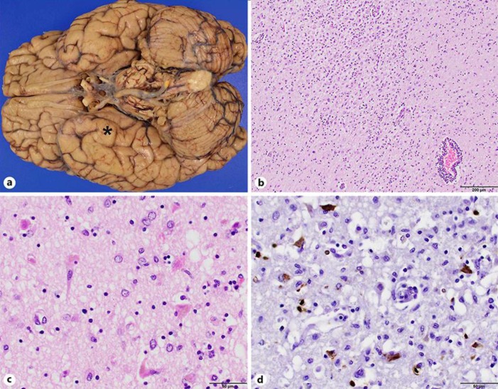
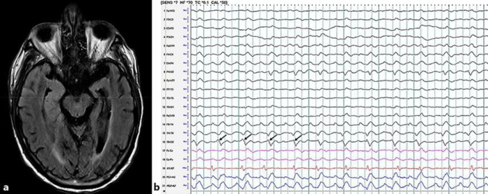
An 88-year-old man presented with a 1-month history of altered mental status and seizures. His electrographic and imaging findings were suggestive of herpes simplex encephalitis (HSE), for which he was empirically treated with acyclovir. He underwent two lumbar punctures 3 days apart; both cerebrospinal fluid analyses tested negative for herpes simplex virus (HSV) by polymerase chain reaction (PCR). These negative results and his continued deterioration after 9 days of acyclovir therapy prompted treatment with steroids for possible autoimmune encephalitis. Shortly after the change in management, the patient died from cardiac arrest. At autopsy, his brain showed both gross and microscopic evidence of encephalitis and was positive for HSV by immunohistochemistry. This fatal case of HSE emphasizes the limitations of HSV PCR and the importance of clinical suspicion in the diagnosis and management of this disease.
Keywords: Encephalitis; False-negative results; Herpes simplex virus; Polymerase chain reaction.
Figures
References
-
- Jakob NJ, Lenhard T, Schnitzler P, Rohde S, Ringleb PA, Steiner T, et al. Herpes simplex virus encephalitis despite normal cell count in the cerebrospinal fluid. Crit Care Med. 2012 Apr;40((4)):1304–8. - PubMed
-
- Denes E, Labach C, Durox H, et al. Intrathecal synthesis of specific antibodies as a marker of herpes simplex encephalitis in patients with negative PCR. Swiss Med Wkly. 2010;140 w13107. - PubMed
-
- DeBiasi RL, Kleinschmidt-DeMasters BK, Richardson-Burns S, Tyler KL. Central nervous system apoptosis in human herpes simplex virus and cytomegalovirus encephalitis. J Infect Dis. 2002 Dec;186((11)):1547–57. - PubMed
Publication types
LinkOut - more resources
Full Text Sources
